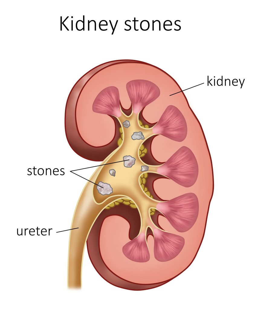
Urolithiasis, commonly referred to as urinary stone disease, denotes the formation of calculi (stones) within the urinary tract, including the kidneys, ureters, bladder, or urethra. These stones form due to supersaturation of minerals and salts, leading to crystallization and aggregation. It remains a globally prevalent urological disorder with significant recurrence rates and clinical burden.
Classification of Urinary Stones
Urinary calculi are categorized based on their chemical composition:
| Stone Type | Composition | Associated Conditions |
|---|---|---|
| Calcium Oxalate | Calcium + Oxalate | Hypercalciuria, dehydration |
| Calcium Phosphate | Calcium + Phosphate (apatite/brushite) | Renal tubular acidosis, alkaline urine |
| Uric Acid | Uric acid crystals | Gout, low urine pH |
| Struvite | Magnesium ammonium phosphate | Chronic UTIs with urease-producing bacteria |
| Cystine | Cystine amino acid | Genetic disorder (cystinuria) |
Pathophysiology of Stone Formation
Stone formation follows a multistep process driven by biochemical and anatomical abnormalities. The supersaturation of urine with insoluble compounds initiates crystallization, followed by crystal retention and growth, especially at sites of urinary stasis or obstruction.
Risk Factors for Urolithiasis
Modifiable Risk Factors
- Low fluid intake: Reduces urine volume and increases solute concentration.
- High oxalate diet: Leafy greens, tea, chocolate.
- Excess animal protein: Promotes uric acid and calcium stones.
- Salt-rich diet: Increases calcium excretion.
Non-Modifiable Risk Factors
- Genetic predisposition
- Male sex
- Age (30–60 years)
- Geographical regions with high temperatures
Clinical Presentation of Urolithiasis
The clinical symptoms of urinary stones vary with their size, location, and movement:
- Renal Colic: Sudden, severe flank pain radiating to the groin.
- Hematuria: Microscopic or visible blood in urine.
- Dysuria: Pain during urination, especially with lower tract stones.
- Nausea and vomiting
- Fever and chills: Indicate secondary infection, warrant urgent care.
Diagnostic Evaluation
1. Imaging Studies
| Modality | Utility |
|---|---|
| Non-contrast CT KUB | Gold standard for detection and localization |
| Ultrasound | First-line for pregnant patients or children |
| X-ray KUB | Detects radiopaque stones (calcium, struvite) |
| IV Urography | Evaluates function and obstruction |
2. Laboratory Tests
- Urinalysis: Detects crystals, hematuria, pyuria.
- 24-hour urine collection: Measures stone-forming constituents.
- Serum tests: Calcium, uric acid, creatinine levels.
Management of Urolithiasis
Conservative Medical Management
Indicated for stones <5 mm with minimal symptoms:
- Increased fluid intake: At least 2.5–3 liters/day
- Analgesia: NSAIDs or opioids for pain control
- Alpha-blockers (e.g., tamsulosin): Facilitate ureteral stone passage
Pharmacological Therapy
- Potassium citrate: Alkalinizes urine, used in uric acid or cystine stones
- Thiazide diuretics: Reduce urinary calcium excretion
- Allopurinol: Lowers uric acid levels in hyperuricemia
Surgical and Minimally Invasive Interventions
| Procedure | Indication |
|---|---|
| ESWL (Shock Wave Lithotripsy) | Stones <2 cm, non-cystine, radiopaque |
| URS (Ureteroscopy) | Ureteral or renal pelvic stones |
| PCNL (Percutaneous Nephrolithotomy) | Large stones >2 cm or staghorn calculi |
| Open/Laparoscopic Surgery | Rare, for complex or failed minimally invasive cases |
Complications of Untreated Stones
- Hydronephrosis: Backpressure causes renal dilation
- Infection or sepsis
- Loss of renal function
- Ureteral stricture
- Recurrent stones: Up to 50% recurrence in 5 years
Prevention Strategies for Urolithiasis
Hydration and Dietary Modifications
- Maintain urine output >2.5 L/day
- Limit dietary oxalates and sodium
- Moderate animal protein intake
- Increase intake of citrate-rich fruits (lemon, orange)
Pharmacologic Prevention
| Condition | Preventive Agent |
|---|---|
| Hypercalciuria | Thiazides |
| Hypocitraturia | Potassium citrate |
| Hyperuricosuria | Allopurinol |
| Cystinuria | Tiopronin, D-penicillamine |
Prognosis and Long-Term Monitoring
With early diagnosis and appropriate intervention, the prognosis of urolithiasis is favorable. Regular follow-up with imaging and 24-hour urine analysis is essential for recurrence prevention. Long-term lifestyle adjustments significantly reduce the stone burden and improve renal outcomes.
Frequently Asked Questions:
What are the most common types of kidney stones?
Calcium oxalate stones are the most prevalent, followed by uric acid, struvite, and cystine stones.
Can small kidney stones pass without treatment?
Yes, stones <5 mm often pass spontaneously with adequate hydration and medical support.
How can diet influence kidney stone formation?
High oxalate, sodium, and animal protein increase stone risk; citrate-rich and plant-based diets are protective.
Is urolithiasis a recurring condition?
Yes, recurrence is common without preventive measures, with up to 80% recurrence over 10 years.
Can drinking lemon water help prevent stones?
Yes, citrate in lemon juice inhibits stone formation and promotes urinary alkalinity.