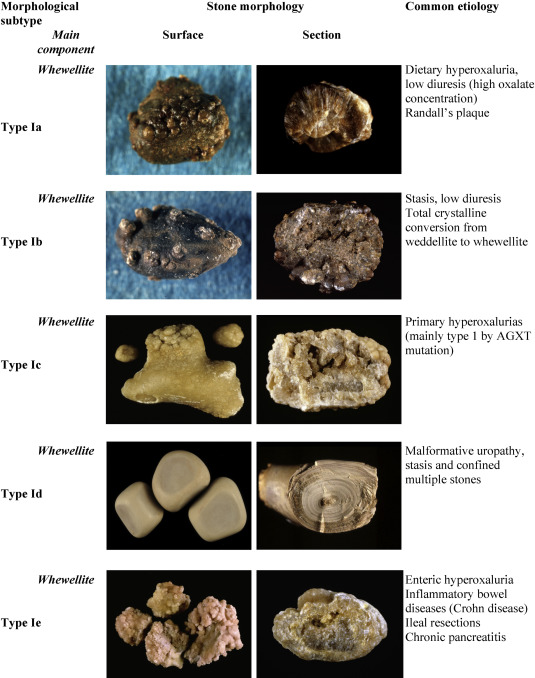
Urate renal calculi, commonly referred to as uric acid kidney stones, are crystalline deposits formed within the renal system due to elevated uric acid concentration in the urine. These stones are typically radiolucent, acid-soluble, and frequently associated with persistently acidic urinary pH, hyperuricemia, and increased purine metabolism. Effective management of urate calculi involves precise diagnosis, pharmacologic and dietary interventions, and consistent monitoring to prevent recurrence.

Pathogenesis of Urate Stone Formation
Urate stones form when there is a supersaturation of uric acid in acidic urine, usually with a pH below 5.5. Unlike calcium stones, urate stones do not bind with oxalate or phosphate but precipitate directly in acidic environments. This process is significantly influenced by urinary pH, urine volume, and uric acid concentration.
Etiological Factors and Risk Profile
Primary Risk Factors:
- Hyperuricosuria: Excessive urinary excretion of uric acid (>800 mg/day in men, >750 mg/day in women)
- Persistently Acidic Urine: pH < 5.5 enhances uric acid crystallization
- Low Urine Volume: Dehydration or inadequate fluid intake reduces solubility
- High Purine Diet: Organ meats, red meats, seafood, and alcohol elevate uric acid levels
- Metabolic Syndrome: Obesity, insulin resistance, and type 2 diabetes correlate with acidic urine
- Genetic Predisposition: Certain inherited disorders of purine metabolism
Clinical Presentation of Uric Acid Nephrolithiasis
Urate renal calculi may be asymptomatic or present with acute symptoms depending on stone size and location.
Common Symptoms:
- Severe flank or abdominal pain (renal colic)
- Hematuria (microscopic or gross)
- Nausea and vomiting
- Dysuria and urinary urgency
- Urinary tract infections in obstructive cases
In some cases, bilateral urate stones may result in acute obstructive uropathy, leading to renal impairment.
Diagnostic Evaluation
Because uric acid stones are radiolucent, standard abdominal X-rays may fail to detect them. Diagnostic precision requires a combination of imaging and biochemical evaluation.
Key Diagnostic Tools:
- Non-Contrast CT Scan: Gold standard for detecting radiolucent urate stones
- Ultrasound: Useful in identifying hydronephrosis or large obstructive stones
- Urinalysis: Reveals acidic urine, hematuria, and crystals
- 24-Hour Urine Analysis: Quantifies uric acid, pH, and other lithogenic factors
- Serum Uric Acid Levels: May be elevated, but not always diagnostic
Management of Urate Renal Calculi
Pharmacological Therapy:
- Urine Alkalinization (Primary Strategy):
- Potassium Citrate: 20–40 mEq/day to maintain pH between 6.0–6.5
- Sodium Bicarbonate: Alternative option, less preferred due to sodium load
- Xanthine Oxidase Inhibitors:
- Allopurinol: 100–300 mg/day for patients with hyperuricemia or recurrent stones
- Febuxostat: For patients intolerant to allopurinol
- Hydration:
- Maintain urine output >2.5 L/day
- Encourage low-sodium, high-fluid intake
Dietary Modification:
- Limit Purine Intake: Avoid organ meats, shellfish, and red meat
- Increase Fruits and Vegetables: Promote urine alkalinity
- Reduce Fructose and Alcohol Consumption: Lowers endogenous uric acid production
Interventional Approaches
In cases of large or obstructive stones, surgical or minimally invasive techniques may be required.
Options Include:
- Extracorporeal Shock Wave Lithotripsy (ESWL): Less effective due to stone composition
- Ureteroscopy with Laser Lithotripsy: Effective for mid and distal ureteric stones
- Percutaneous Nephrolithotomy (PCNL): Used for large or complex renal stones
Prompt decompression is critical in obstructive uropathy to prevent renal failure.
Prevention of Recurrence
Preventive strategies must be long-term and tailored to individual metabolic profiles.
Key Preventive Measures:
- Maintain Urine pH Between 6.0–6.5
- Consistent Hydration and Urine Dilution
- Ongoing Dietary Adjustments
- Regular Follow-Up with 24-Hour Urine Testing
- Management of Comorbid Conditions such as diabetes and obesity
Comparison with Other Renal Calculi Types
| Feature | Uric Acid Stones | Calcium Oxalate Stones | Struvite Stones |
|---|---|---|---|
| Radiographic Appearance | Radiolucent (X-ray invisible) | Radiopaque (visible) | Radiopaque |
| Urine pH | Acidic (<5.5) | Variable | Alkaline (>7.0) |
| Composition | Uric acid | Calcium oxalate | Magnesium ammonium phosphate |
| Associated Conditions | Gout, Metabolic Syndrome | Hypercalciuria, Hyperoxaluria | UTIs with urease-producing bacteria |
Urate renal calculi are a distinct form of kidney stone disease associated with acidic urinary pH, hyperuricosuria, and dietary factors. Effective management depends on prompt identification through advanced imaging, urine alkalization, pharmacologic interventions, and lifestyle modifications. Long-term success hinges on preventive strategies including pH regulation, hydration, and dietary restraint. Given their unique metabolic profile and radiolucency, urate stones require tailored diagnostic and therapeutic approaches.

