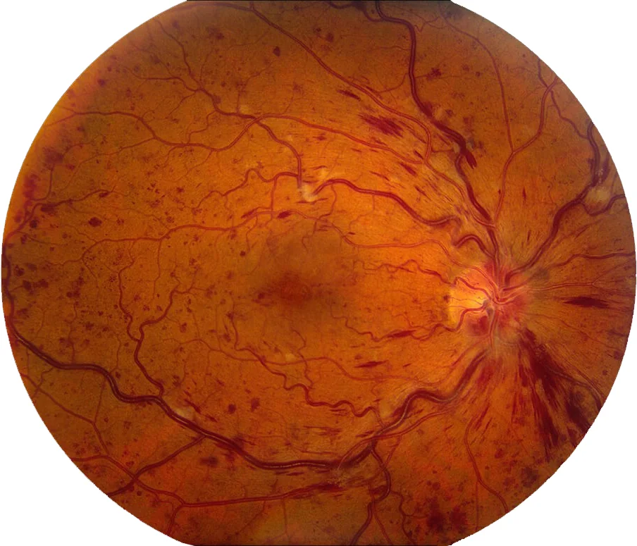
Central retinal vein occlusion with macular edema (CRVO) is a significant retinal vascular disorder characterized by the obstruction of the central retinal vein, leading to retinal damage. A frequent complication of CRVO is macular edema, which results in swelling of the macula due to fluid accumulation. This combination can severely impair vision, making early diagnosis and effective management critical. This guide provides an in-depth exploration of CRVO with macular edema, focusing on causes, symptoms, diagnostic methods, and treatment options.

Understanding Central Retinal Vein Occlusion
CRVO occurs when the central retinal vein, responsible for draining blood from the retina, becomes blocked. This blockage can result in increased venous pressure, hemorrhages, and fluid leakage into the retina, ultimately leading to macular edema.
Types of CRVO
- Non-Ischemic CRVO: A milder form with partial venous blockage and relatively better visual prognosis.
- Ischemic CRVO: A severe form with complete venous blockage, higher risk of complications, and poorer visual outcomes.
Risk Factors
- Systemic Conditions: Hypertension, diabetes mellitus, and hyperlipidemia.
- Ophthalmic Conditions: Glaucoma and optic disc abnormalities.
- Lifestyle Factors: Smoking and sedentary behavior.
- Hematologic Disorders: Hypercoagulable states.
Macular Edema in CRVO
Macular edema occurs when fluid leaks from damaged retinal blood vessels, causing the macula to swell. This swelling distorts central vision, significantly affecting daily activities like reading and recognizing faces.
Symptoms
- Blurred or distorted central vision.
- Reduced color perception.
- Central scotoma (a dark or blank spot in vision).
Diagnosis of CRVO with Macular Edema
Clinical Examination
- Visual Acuity Testing: To assess the degree of vision loss.
- Ophthalmoscopy: To identify retinal hemorrhages, venous engorgement, and macular swelling.
Diagnostic Imaging
- Fluorescein Angiography (FA): Highlights vascular leakage and ischemia.
- Optical Coherence Tomography (OCT): Provides high-resolution images of the retina to quantify macular edema.
- OCT Angiography: Non-invasive imaging to assess retinal vasculature.
Treatment Options
Intravitreal Injections
- Anti-VEGF Therapy
- Medications: Ranibizumab, Aflibercept, Bevacizumab.
- Mechanism: Inhibits vascular endothelial growth factor (VEGF) to reduce fluid leakage and neovascularization.
- Frequency: Monthly injections, gradually reduced based on response.
- Corticosteroids
- Medications: Dexamethasone implants (Ozurdex), Triamcinolone acetonide.
- Benefits: Reduces inflammation and vascular permeability.
- Considerations: Increased risk of cataracts and glaucoma.
Laser Photocoagulation
- Indicated for non-responsive macular edema or ischemic CRVO with neovascularization.
- Mechanism: Reduces retinal ischemia and prevents further complications.
Systemic Management
- Optimizing control of underlying systemic conditions like hypertension and diabetes.
- Anticoagulation therapy may be considered in specific cases.
Complications and Prognosis
- Complications: Neovascular glaucoma, retinal detachment, and persistent vision loss.
- Prognosis: Varies based on the type of CRVO, extent of macular edema, and promptness of treatment. Non-ischemic CRVO has a better prognosis compared to ischemic CRVO.
Prevention Strategies
- Regular Eye Exams: Essential for early detection in high-risk individuals.
- Manage Systemic Conditions: Strict control of blood pressure, blood sugar, and lipid levels.
- Healthy Lifestyle: Avoid smoking, maintain a balanced diet, and engage in regular exercise
MYHEALTHMAG