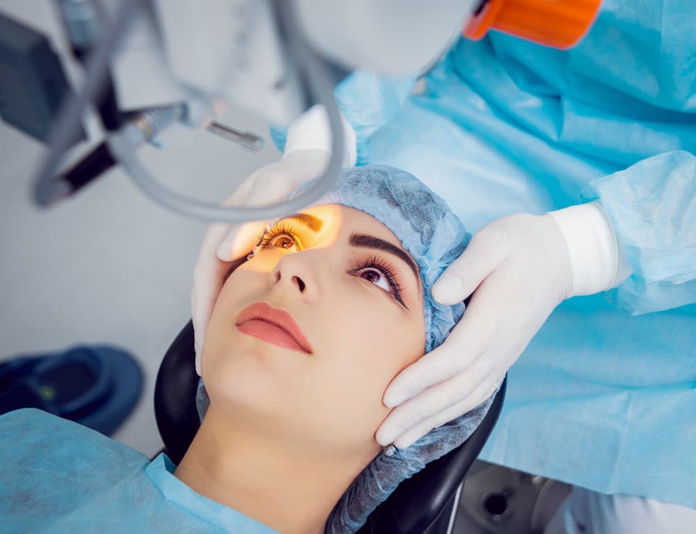
Cataract surgery (cataract surgery adjunct to enhance visualization) is one of the most commonly performed procedures worldwide, with advancements continually refining surgical outcomes. A key aspect of achieving optimal results lies in enhanced visualization during surgery, which allows surgeons to perform precise, effective, and safe operations. Various adjuncts and innovations have emerged to address this need. This article explores the cutting-edge tools and techniques used to improve visualization in cataract surgery, ensuring superior patient outcomes.

Importance of cataract surgery adjunct to enhance visualization
Visualization plays a pivotal role in cataract surgery, as the success of the procedure depends on the surgeon’s ability to clearly see intraocular structures. Poor visibility can increase risks, such as incomplete lens removal, damage to adjacent tissues, or improper placement of intraocular lenses (IOLs). Enhanced visualization minimizes these risks and ensures:
- Improved surgical precision
- Shortened procedure time
- Reduced postoperative complications
- Enhanced patient safety and satisfaction
Adjuncts to Enhance Visualization in Cataract Surgery
1. Ophthalmic Viscosurgical Devices (OVDs)
OVDs play a critical role in maintaining space and protecting ocular structures during surgery. Their benefits include:
- Maintaining the anterior chamber depth.
- Coating and protecting corneal endothelium from mechanical damage.
- Facilitating capsulorhexis by stabilizing the capsule.
Commonly used OVDs include cohesive, dispersive, and viscoadaptive agents, each chosen based on the surgical phase.
2. Advanced Intraoperative Microscopes
State-of-the-art surgical microscopes enhance the surgeon’s ability to visualize fine details of intraocular structures. Features include:
- High-definition optics for detailed imaging.
- Integrated heads-up displays.
- Digital recording capabilities for training and review.
- Real-time contrast enhancement using optical coherence tomography (OCT).
3. Intraoperative OCT (iOCT)
The integration of OCT technology provides real-time cross-sectional imaging of ocular tissues during surgery. Benefits of iOCT include:
- Enhanced depth perception.
- Real-time guidance during challenging cases, such as those with dense cataracts.
- Improved visualization of posterior capsule integrity.
4. Capsular Dyes
Capsular dyes improve the visibility of transparent or faint structures like the anterior capsule. Common dyes include:
- Trypan Blue: Most widely used for staining the anterior capsule.
- Brilliant Blue: Primarily used for retinal and vitreous visualization.
- Indocyanine Green (ICG): Occasionally used in complicated cases.
5. Digital Visualization Systems
Heads-up displays and 3D digital visualization systems provide enhanced depth perception and ergonomics for the surgeon. Key advantages include:
- Magnified, high-resolution 3D imaging.
- Reduced surgeon fatigue due to ergonomic designs.
- Compatibility with advanced imaging systems for better outcomes.
6. Improved Surgical Instruments
High-quality surgical instruments designed with precision optics aid in:
- Precise incision placement.
- Improved phacoemulsification efficiency.
- Reduced risk of tissue damage.
Innovations in Visualization Techniques
Femtosecond Laser-Assisted Cataract Surgery (FLACS)
FLACS has revolutionized cataract surgery by automating critical steps with laser precision. Enhanced visualization benefits include:
- Better delineation of lens fragments.
- Improved capsulorhexis accuracy.
- Reduced phaco energy required, minimizing endothelial cell loss.
Augmented Reality (AR) Integration
AR systems superimpose real-time digital information over the surgical field, allowing surgeons to:
- Navigate with increased precision.
- Identify ocular structures more effectively.
- Plan and execute complex maneuvers with confidence.
Challenges and Future Directions
Despite these advancements, several challenges remain:
- Cost: High-end visualization systems can be expensive, limiting accessibility in resource-poor settings.
- Learning Curve: Adopting new technologies requires time and training.
- Customization: Tailoring adjuncts to individual patient needs remains a challenge.
Future innovations aim to bridge these gaps by:
- Developing affordable yet effective visualization tools.
- Leveraging AI to provide real-time surgical guidance.
- Enhancing telemedicine capabilities for remote assistance.

