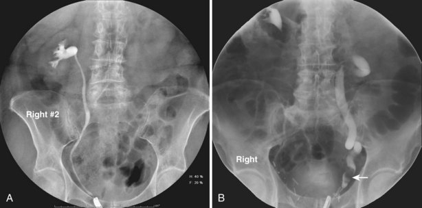
Subcutaneous urography is a valuable imaging technique used to evaluate the urinary system, primarily focusing on the kidneys, ureters, and bladder. In modern radiology, enhancing the contrast resorption process has proven essential for improving diagnostic outcomes. The use of adjuncts for contrast resorption in subcutaneous urography plays a pivotal role in achieving better visualization, facilitating accurate diagnosis, and ensuring the effective assessment of renal function. This technique is critical for clinicians to assess various conditions, such as urinary tract obstructions, kidney stones, and tumors. Understanding the mechanisms, benefits, and advancements in this area can help healthcare professionals make informed decisions regarding diagnostic procedures and treatment plans.

What Is Subcutaneous Urography?
Subcutaneous urography is a diagnostic imaging procedure that involves injecting a contrast medium into the subcutaneous tissues to enhance the visibility of the urinary system during radiographic imaging. Unlike traditional intravenous urography, which uses intravenous contrast agents to highlight the urinary tract, subcutaneous urography offers several advantages, particularly when there are contraindications to intravenous contrast media.
This technique allows for better visualization of anatomical structures such as the kidneys, ureters, and bladder, making it a crucial tool for diagnosing various conditions affecting the urinary system. Additionally, subcutaneous urography has become particularly important for patients with renal dysfunction, those with allergies to iodine-based contrast agents, or individuals with compromised venous access.
The Role of Contrast Resorption in Subcutaneous Urography
Contrast resorption refers to the absorption and distribution of the injected contrast medium within the body. In subcutaneous urography, the contrast agent is injected into the subcutaneous tissue, and it is slowly absorbed into the circulatory system. This process facilitates the contrast medium’s eventual delivery to the kidneys, where it is filtered and excreted, providing clear imaging of the urinary tract.
The success of subcutaneous urography largely depends on the effective resorption and distribution of the contrast material. Enhancing contrast resorption can improve imaging quality, enabling more accurate evaluations of kidney function and the detection of abnormalities in the urinary system. An efficient contrast resorption process helps ensure that the contrast medium reaches the renal parenchyma, providing enhanced clarity of the kidneys, bladder, and ureters.
Methods to Enhance Contrast Resorption in Subcutaneous Urography
To optimize the resorption and imaging quality of subcutaneous urography, several adjuncts and techniques can be employed. These methods help ensure that the contrast agent is absorbed efficiently, allowing for better delineation of the urinary structures.
1. Hydration and Fluid Management
Adequate hydration before and after the procedure is one of the key strategies to promote effective contrast resorption. Patients are often instructed to drink sufficient fluids to ensure optimal renal perfusion. This can facilitate the movement of the contrast agent through the body, improving its distribution and clearance by the kidneys.
Additionally, ensuring that the patient remains well-hydrated helps minimize the risk of nephrotoxicity, which can occasionally occur when contrast agents are not properly eliminated. Proper fluid management also prevents dehydration, a common risk factor that can affect renal function and compromise the imaging process.
2. Temperature Regulation of Contrast Medium
The temperature of the contrast medium has been shown to influence its resorption and effectiveness. Studies suggest that warming the contrast medium before injection can accelerate its absorption, making the imaging process more efficient. By reducing the viscosity of the contrast agent, it moves more easily through the subcutaneous tissues and is absorbed more rapidly by the body, improving the overall imaging quality.
This technique also minimizes patient discomfort, as cold contrast media can sometimes cause pain or a burning sensation during injection. By warming the contrast medium, clinicians can improve patient experience while optimizing diagnostic outcomes.
3. Choice of Contrast Medium
The selection of an appropriate contrast medium is crucial in subcutaneous urography. Certain contrast agents are specifically designed to enhance absorption and distribution within the body. For example, low-osmolar and iso-osmolar contrast agents are preferred for their ability to be absorbed efficiently and reduce the risk of adverse reactions. These agents facilitate better visualization of the urinary tract by providing clearer images and ensuring that the contrast medium is appropriately resorbed.
Furthermore, advancements in contrast media, such as the use of newer iodine-based agents, have enhanced imaging capabilities. These agents are specifically formulated to offer improved contrast retention in the tissues, which is essential for high-quality imaging.
4. Massage and Physical Manipulation
In some cases, manual manipulation of the injection site or gentle massage of the affected area may be recommended to promote the even distribution of the contrast medium. This technique can help the contrast agent to spread more evenly across the subcutaneous tissue, ensuring that it reaches the target areas efficiently. While not always necessary, this adjunct can be particularly helpful in ensuring that the contrast material is absorbed effectively and in a timely manner.
Clinical Applications of Subcutaneous Urography Adjuncts for Contrast Resorption
Subcutaneous urography adjuncts for contrast resorption have a wide range of clinical applications, particularly in diagnostic scenarios where traditional imaging methods may not be feasible or sufficient.
1. Renal Function Assessment
One of the primary uses of subcutaneous urography is to evaluate renal function and detect abnormalities such as renal obstructions, kidney stones, or tumors. By enhancing the resorption of the contrast medium, clinicians can obtain clearer images of the kidneys, allowing for more precise assessments of kidney size, shape, and function.
This technique is particularly useful in patients with compromised renal function, where intravenous contrast may be contraindicated. By injecting the contrast medium subcutaneously, healthcare providers can still obtain essential diagnostic information without placing the patient at risk for nephrotoxicity.
2. Obstructive Uropathy Diagnosis
Subcutaneous urography is invaluable in diagnosing conditions like obstructive uropathy, where a blockage in the urinary tract prevents the normal flow of urine. The enhanced contrast resorption process enables healthcare professionals to observe the dilation of the renal pelvis or ureters, which is indicative of obstruction. This helps guide treatment decisions and identify the most appropriate interventions, whether surgical or medical.
3. Urinary Tract Infections (UTIs)
In cases of chronic or recurrent urinary tract infections (UTIs), subcutaneous urography can help identify anatomical abnormalities that may predispose patients to infections. By improving the quality of imaging through better contrast resorption, clinicians can detect conditions such as vesicoureteral reflux or congenital anomalies that may contribute to UTIs.
4. Tumor Detection and Staging
Subcutaneous urography plays a critical role in the detection and staging of urinary tract tumors. The enhanced imaging capabilities provided by contrast resorption allow for better visualization of tumors within the kidneys, ureters, and bladder. This is especially important for assessing the size, location, and extent of tumors, which is essential for determining the most effective treatment plan.
Future Directions in Subcutaneous Urography and Contrast Resorption
The field of subcutaneous urography and contrast resorption continues to evolve, with ongoing research aimed at improving the techniques and agents used for imaging.
1. Advancements in Contrast Media
Newer generations of contrast media are being developed to provide even better imaging results with fewer side effects. These next-generation agents are designed to be more efficient in terms of absorption and resorption, offering improved image quality and reducing the likelihood of adverse reactions.
2. Integration with Other Imaging Modalities
As technology advances, subcutaneous urography is increasingly being combined with other imaging techniques such as ultrasound, MRI, and CT scans. This multimodal approach enhances the overall diagnostic capabilities, providing a more comprehensive assessment of the urinary system.
3. Personalized Medicine and Tailored Imaging Protocols
In the future, imaging protocols for subcutaneous urography may become more personalized, taking into account individual patient characteristics such as age, renal function, and medical history. This approach will allow for the development of tailored contrast resorption strategies that optimize diagnostic accuracy and minimize risks.
Subcutaneous urography adjuncts for contrast resorption represent a crucial advancement in diagnostic imaging, particularly for patients with specific contraindications or complications. By improving the resorption of contrast media, healthcare professionals can obtain clearer, more accurate images of the urinary system, aiding in the diagnosis and management of various renal and urological conditions. With continued advancements in contrast media and imaging techniques, the future of subcutaneous urography promises to enhance diagnostic precision and patient outcomes.

