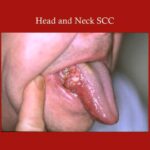
Squamous cell carcinoma of the lung is a common subtype of non-small cell lung cancer (NSCLC), typically originating in the central bronchi and strongly associated with tobacco smoking. As one of the major histological types of lung cancer, it accounts for approximately 20–30% of all NSCLC cases. Timely diagnosis and appropriate staging are essential for effective management.
What Is Squamous Cell Carcinoma of the Lung?
Squamous cell carcinoma (SCC) arises from the squamous epithelial cells that line the airways. These cells undergo malignant transformation, often due to chronic irritation from inhaled carcinogens like tobacco smoke. SCC is typically found in the central regions of the lungs, especially in the larger bronchi.
Major Risk Factors for Pulmonary Squamous Cell Carcinoma
Tobacco Use
The primary risk factor for lung SCC is long-term cigarette smoking. A direct dose-response relationship exists between the number of pack-years and cancer risk.
Environmental Exposure
- Occupational Hazards: Asbestos, silica dust, diesel exhaust.
- Radon Gas: Second-leading cause of lung cancer in nonsmokers.
- Air Pollution: PM2.5 exposure contributes to increased incidence.
Genetic and Molecular Alterations
- TP53 mutations
- EGFR wild-type status (common in SCC)
- Amplification of FGFR1, SOX2, and PIK3CA genes
Clinical Symptoms of Squamous Cell Carcinoma of the Lung
Most patients present with symptoms due to central airway obstruction or local invasion:
- Persistent cough
- Hemoptysis (coughing up blood)
- Chest pain or tightness
- Wheezing and shortness of breath
- Recurrent respiratory infections
- Hoarseness (from recurrent laryngeal nerve involvement)
- Unintended weight loss and fatigue
Advanced disease may cause superior vena cava syndrome, pleural effusion, or bone pain due to metastases.
Diagnostic Evaluation of Lung SCC
Imaging Studies
- Chest X-ray: May reveal hilar mass or atelectasis
- CT Scan: Essential for determining tumor size, location, and nodal involvement
- PET-CT: Used for staging and detecting metastasis
Bronchoscopy with Biopsy
Provides direct visualization and tissue sampling of central tumors.
Histopathological Examination
- Keratinization and intercellular bridges are hallmark features.
- Immunohistochemistry: Positive for p40, CK5/6, and p63
Molecular Testing
- Unlike adenocarcinoma, SCC is usually negative for EGFR or ALK mutations.
- Testing for PD-L1 expression is critical for immunotherapy eligibility.
TNM Staging of Lung Squamous Cell Carcinoma
Staging follows the AJCC 8th Edition TNM classification, which determines prognosis and therapeutic decisions.
| TNM Component | Description |
|---|---|
| T (Tumor) | Size and extent of primary tumor |
| N (Nodes) | Regional lymph node involvement |
| M (Metastasis) | Presence of distant metastatic disease |
Stage Grouping Example:
- Stage I: Localized tumor without nodal involvement
- Stage II: Larger tumor or involvement of nearby lymph nodes
- Stage III: Spread to mediastinal nodes or nearby structures
- Stage IV: Distant metastasis (brain, liver, bones)
Treatment Options for Lung Squamous Cell Carcinoma
Surgical Resection
Indicated for early-stage, operable tumors:
- Lobectomy: Preferred surgical approach
- Pneumonectomy: For centrally located extensive tumors
Radiation Therapy
Used as:
- Adjuvant post-surgery in high-risk cases
- Definitive in unresectable disease or medically inoperable patients
Chemotherapy
Platinum-based doublets (cisplatin/carboplatin with gemcitabine or paclitaxel) are standard for:
- Stage III unresectable tumors
- Stage IV metastatic disease
Immunotherapy
Checkpoint inhibitors such as nivolumab, pembrolizumab, and atezolizumab are now first-line or second-line therapies depending on PD-L1 expression and previous treatment.
Targeted Therapy
Less applicable in SCC due to lack of common driver mutations, though FGFR inhibitors are under investigation in select cases.
Prognosis and Survival Statistics
Prognosis is influenced by stage, performance status, molecular profile, and treatment response.
| Stage | 5-Year Survival Rate |
|---|---|
| Stage I | 45–60% |
| Stage II | 30–40% |
| Stage III | 10–25% |
| Stage IV | <5–10% |
HPV-positive SCC of the lung is extremely rare and not prognostically relevant as it is in head and neck cancers.
Prevention and Risk Reduction Strategies
- Smoking Cessation: Most effective strategy to reduce incidence
- Radon Testing: Especially in residential areas
- Occupational Safety: Use of personal protective equipment
- Early Screening: Low-dose CT scan for high-risk individuals aged 50–80 with ≥20 pack-year smoking history
Frequently Asked Questions:
Is squamous cell carcinoma of the lung aggressive?
Yes. It often presents late and progresses rapidly, especially when centrally located and undetected early.
Can lung SCC be cured?
Early-stage SCC can be curable with surgery or combined modality therapy. Advanced cases are typically managed palliatively.
How is SCC different from adenocarcinoma?
SCC is centrally located and strongly smoking-related; adenocarcinoma typically arises peripherally and may occur in nonsmokers.
Is immunotherapy effective in SCC?
Yes. Immunotherapy improves survival in advanced SCC, particularly when PD-L1 expression is high.
Is there a screening test for lung SCC?
Low-dose CT screening is recommended for high-risk individuals, which can help detect early-stage lung cancers including SCC.
Squamous cell carcinoma of the lung remains a significant global health burden, largely driven by tobacco use. Early recognition, precise staging, and personalized treatment strategies including surgery, radiation, chemotherapy, and immunotherapy are crucial for improving outcomes. As research evolves, molecular profiling and novel agents continue to refine care pathways for this aggressive malignancy.