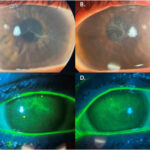
Punctate keratitis is a clinical condition marked by inflammation of the cornea, specifically presenting as multiple small epithelial erosions or opacities. These punctate lesions are most commonly seen on the anterior corneal surface and are detectable through slit-lamp biomicroscopy and fluorescein staining. Although often benign, punctate keratitis can significantly affect visual clarity and ocular comfort, and its presence typically signals underlying ocular surface pathology.
Etiopathogenesis of Punctate Keratitis
Disruption of the Corneal Epithelium
The corneal epithelium acts as a barrier protecting the eye from external insults. Punctate keratitis occurs when this protective layer is compromised due to mechanical, chemical, infectious, or inflammatory causes.
Common Causative Factors
- Dry Eye Syndrome: Tear film instability and hyperosmolarity damage epithelial cells.
- Contact Lens Use: Friction and hypoxia-induced epithelial stress.
- Blepharitis: Chronic eyelid inflammation causing tear film abnormalities.
- Toxicity: Long-term use of preservatives like benzalkonium chloride in eye drops.
- Ultraviolet Exposure: Solar or artificial UV rays causing photokeratitis.
- Infections: Viral (herpes simplex, adenovirus), bacterial, or fungal keratitis.
- Autoimmune Diseases: Conditions like Sjögren’s syndrome or ocular cicatricial pemphigoid.
Clinical Features and Symptomatology
Patients with punctate keratitis may present with a wide range of symptoms that vary based on severity and the underlying cause.
Symptoms Include:
- Foreign body sensation
- Photophobia
- Tearing or watery eyes
- Ocular redness
- Burning or stinging
- Visual fluctuations or blurriness
In severe cases, visual acuity may be temporarily diminished due to epithelial irregularities or associated stromal involvement.
Diagnostic Techniques for Accurate Evaluation
Timely and accurate diagnosis of punctate keratitis is essential for effective treatment. A detailed history and slit-lamp examination remain foundational.
Staining Patterns and Significance
- Inferior Staining: Suggests exposure or blepharitis.
- Interpalpebral Staining: Common in dry eye disease.
- Diffuse Staining: Seen in toxic or allergic reactions.
- Linear/Dendritic Pattern: Indicates herpetic keratitis.
Differential Diagnosis of Punctate Corneal Lesions
Accurate differentiation of punctate keratitis from other corneal conditions ensures appropriate intervention.
| Condition | Distinguishing Features |
|---|---|
| Thygeson’s SPK | Bilateral, chronic, coarse lesions with minimal redness |
| Herpes Simplex Keratitis | Unilateral, dendritic ulcers with terminal bulbs |
| Exposure Keratopathy | Inferior punctate lesions, lagophthalmos |
| Toxic Keratopathy | Diffuse epithelial staining from medications or preservatives |
| Filamentary Keratitis | Mucin filaments on corneal surface in chronic dry eye |
Treatment Modalities for Punctate Keratitis
Effective treatment necessitates addressing both the corneal surface damage and the underlying etiology.
General Therapeutic Measures
- Lubricating Eye Drops: Preservative-free artificial tears to restore tear film.
- Ocular Surface Protection: Eyelid hygiene, humidifiers, protective eyewear.
- Medication Review: Discontinue or replace toxic topical agents.
Etiology-Specific Treatments
| Cause | Treatment Strategy |
|---|---|
| Dry Eye | Cyclosporine, lifitegrast, punctal plugs |
| Infectious | Antivirals (e.g., ganciclovir for HSV), antibiotics if bacterial |
| Blepharitis | Warm compresses, lid scrubs, oral doxycycline |
| Toxic | Cease offending drug, introduce supportive care |
| UV Exposure | Lubrication, cold compresses, anti-inflammatory drops |
Advanced Interventions
- Bandage Contact Lenses: Promote epithelial healing in recurrent erosions.
- Amniotic Membrane Grafts: For severe, persistent epithelial defects.
- Topical Autologous Serum: Contains growth factors and vitamins supporting epithelial regeneration.
Prognosis and Follow-Up
With prompt intervention, punctate keratitis is usually reversible and leaves no permanent damage. However, chronic or improperly managed cases may lead to complications such as:
- Recurrent corneal erosions
- Corneal scarring or haze
- Persistent epithelial defects
- Secondary infections
Patients should be regularly monitored until complete resolution of epithelial lesions and stabilization of the underlying condition.
Preventive Measures and Long-Term Management
Preventing recurrence of punctate keratitis is essential, particularly for patients with chronic ocular surface diseases.
Prevention Strategies
- Consistent use of artificial tears in dry environments
- Avoidance of allergens and irritants
- Strict eyelid hygiene in blepharitis-prone individuals
- Protective eyewear during UV exposure
- Regular ophthalmic reviews for high-risk patients
Punctate keratitis serves as a critical marker of ocular surface compromise, often indicative of deeper underlying pathology. A structured approach to diagnosis and a personalized treatment plan can effectively restore corneal integrity and improve patient quality of life. Emphasis on preventative strategies and long-term care ensures sustainable relief and ocular health preservation.