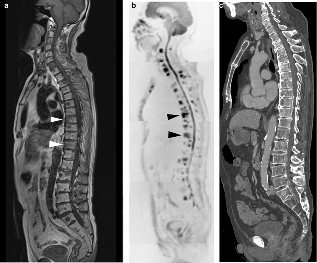
Prostate cancer remains one of the most prevalent malignancies among men globally. Accurate detection of metastatic spread is essential for determining prognosis, guiding therapeutic choices, and evaluating treatment response. Imaging plays a pivotal role in the staging and surveillance of prostatic cancer, particularly in identifying distant metastases. In this comprehensive overview, we examine the latest advances in imaging modalities for prostatic cancer metastases and their clinical applications.

Role of Imaging in Prostatic Cancer Metastases Detection
Importance of Early and Accurate Staging
The progression of prostatic cancer from a localized disease to metastatic involvement significantly alters treatment strategy and outcomes. Early identification of lymphatic, osseous, or visceral metastases enables timely systemic therapy and prevents unnecessary localized interventions.
Imaging is integral in:
- Primary staging of newly diagnosed high-risk patients
- Detection of biochemical recurrence
- Evaluation of treatment response
- Surveillance in advanced disease
PSMA PET/CT: The Gold Standard in Prostate Cancer Imaging
Mechanism of Action and Diagnostic Value
Prostate-Specific Membrane Antigen (PSMA) PET/CT has revolutionized the imaging of prostate cancer metastases. PSMA is a transmembrane glycoprotein significantly overexpressed in prostate cancer cells.
Radiotracers such as 68Ga-PSMA-11 or 18F-DCFPyL bind to PSMA receptors, allowing precise visualization of cancerous tissues even at low PSA levels.
Key advantages include:
- Superior sensitivity and specificity compared to conventional imaging
- Effective in detecting lymph node, bone, and visceral metastases
- Helpful in cases of biochemical recurrence post-prostatectomy or radiotherapy
MRI for Local and Nodal Metastatic Evaluation
Multiparametric MRI (mpMRI)
mpMRI is primarily used for local staging and detecting extracapsular extension or seminal vesicle invasion. It is also valuable in evaluating pelvic lymph nodes and recurrence in the prostatic bed post-treatment.
MRI advantages:
- High-resolution soft tissue contrast
- Useful in characterizing lesions within the prostate gland
- Non-ionizing, repeatable imaging modality
Limitations:
- Less sensitive than PSMA PET/CT for distant metastases
- Limited ability to distinguish small nodal metastases without PSMA targeting
CT and Bone Scintigraphy in Traditional Imaging
Conventional CT
CT scans have historically been used for nodal and visceral staging. However, their limited sensitivity in detecting small lymph nodes or early bone metastases makes them suboptimal compared to newer modalities.
CT is still used in:
- Resource-limited settings
- Complementary evaluation with PET scans
- Emergency assessment of complications (e.g., spinal cord compression)
Bone Scintigraphy (99mTc-MDP)
Bone scintigraphy is used to detect skeletal metastases, which are common in advanced prostate cancer. It identifies areas of increased osteoblastic activity.
Drawbacks:
- Lower specificity—may detect degenerative changes or fractures
- Inability to differentiate active vs treated lesions
- Less accurate in early detection compared to PSMA PET
Whole-Body MRI and Advanced Modalities
Emerging Role of Whole-Body Imaging
Whole-body diffusion-weighted MRI (WB-DWI) is gaining traction as a non-radiative, whole-body alternative for staging and response assessment. It offers:
- Excellent bone marrow lesion detection
- Whole-body coverage without ionizing radiation
- Potential integration in treatment monitoring
Other emerging modalities include:
- PET/MRI hybrid imaging
- Radiomics and AI integration for predictive analytics
Imaging for Biochemical Recurrence
PSA-Triggered Imaging Strategies
Biochemical recurrence is typically indicated by a rising PSA after definitive therapy. Imaging modality selection depends on the absolute PSA level and kinetics.
PSMA PET/CT is favored for:
- PSA levels >0.2 ng/mL post-radical prostatectomy
- PSA doubling time <6 months
- Rising PSA after external beam radiation therapy
Comparative Overview of Imaging Modalities
| Imaging Modality | Best Use Case | Sensitivity | Specificity | Radiation |
|---|---|---|---|---|
| PSMA PET/CT | Metastatic detection & recurrence | High | High | Moderate |
| mpMRI | Local staging & recurrence eval | Moderate | High | None |
| CT | Visceral metastases, emergencies | Low | Moderate | High |
| Bone Scintigraphy | Bone metastasis detection (traditional) | Moderate | Low | Moderate |
| WB-DWI MRI | Whole-body staging & response eval | High | Moderate | None |
Clinical Impact and Treatment Planning
Imaging findings directly influence:
- Determination of curability with localized therapy
- Eligibility for metastasis-directed therapy
- Selection of systemic treatments (androgen deprivation therapy, chemotherapy)
- Prognostication and follow-up planning
Accurate imaging prevents overtreatment or undertreatment and contributes to individualized, patient-centric care.
In the evolving landscape of prostate cancer management, imaging for prostatic cancer metastases has progressed from basic bone scans to highly specific molecular imaging techniques. PSMA PET/CT currently represents the most precise tool for metastatic detection and recurrence evaluation. As technology advances, the integration of functional imaging, AI, and hybrid modalities promises further refinements in the diagnostic pathway. The adoption of these imaging strategies is essential for precision oncology in prostate cancer care.