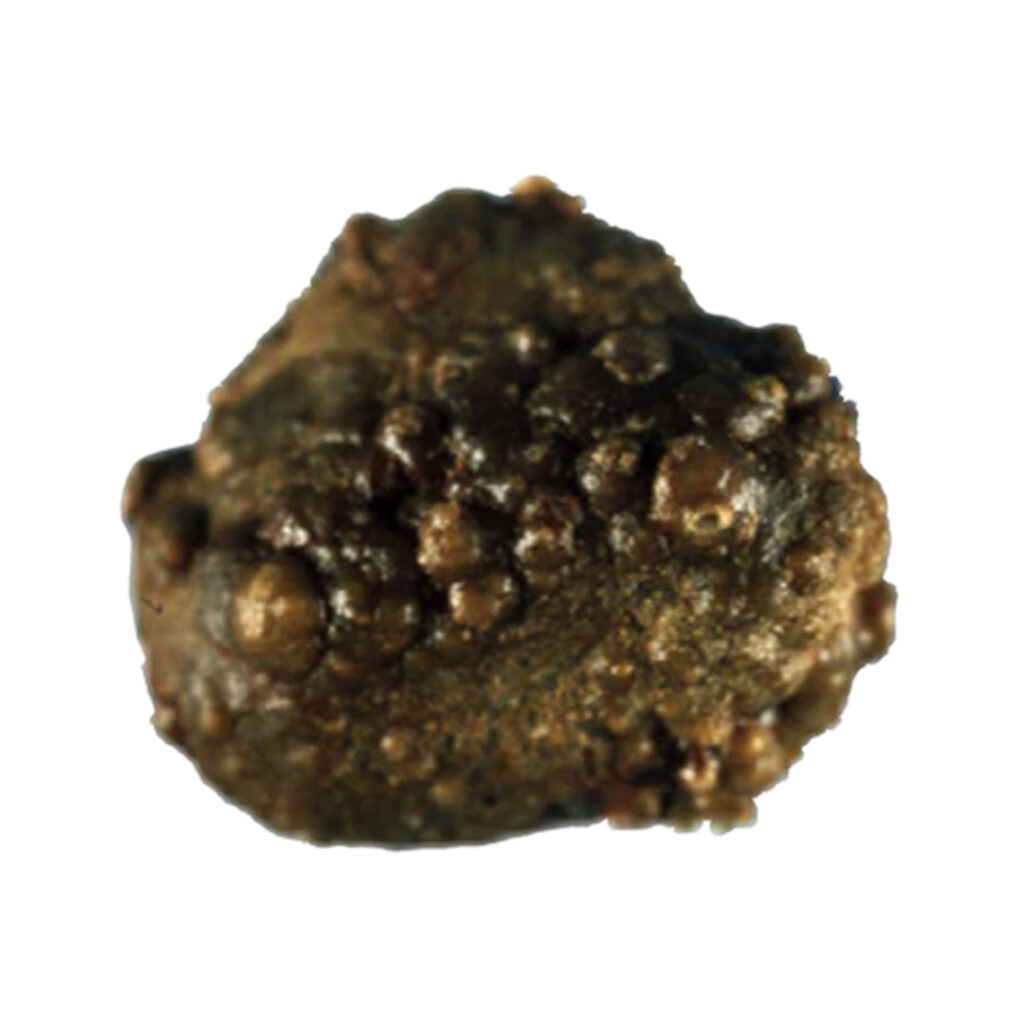
Calcium oxalate renal calculi, commonly referred to as kidney stones, are among the most prevalent types of urinary tract stones, accounting for approximately 70-80% of all cases. These hard mineral deposits form when calcium binds with oxalate in the urine, creating crystalline structures that can lead to significant discomfort and urinary complications. This article explores the causes, symptoms, risk factors, and effective management strategies for calcium oxalate renal calculi.

Pathophysiology of Calcium Oxalate Stones
Calcium oxalate stones form due to supersaturation of calcium and oxalate in the urine. Factors such as low urinary volume, hypercalciuria, hyperoxaluria, and hypocitraturia contribute significantly to stone formation. The aggregation of calcium oxalate crystals within the renal tubules eventually leads to stone development, exacerbated by conditions such as metabolic disorders and chronic dehydration.
Diagram: Formation of Calcium Oxalate Stones
Causes and Risk Factors
Understanding the underlying causes of calcium oxalate renal calculi is crucial for effective prevention and management. Key factors include:
1. Dietary Factors
- High oxalate foods such as spinach, nuts, and chocolate increase urinary oxalate levels.
- Excessive sodium intake promotes calcium excretion in urine, raising the risk of stone formation.
2. Medical Conditions
- Hypercalciuria: Elevated calcium levels in urine.
- Hyperoxaluria: Excess oxalate in urine, often due to dietary or metabolic factors.
- Hypocitraturia: Low citrate levels in urine reduce its ability to inhibit crystal formation.
- Gastrointestinal Disorders: Conditions like Crohn’s disease and gastric bypass surgery can increase oxalate absorption.
3. Genetic Predisposition
Family history plays a significant role, with genetic factors influencing stone formation.
4. Dehydration
Inadequate fluid intake leads to concentrated urine, increasing the likelihood of stone development.
Symptoms
Calcium oxalate renal calculi can remain asymptomatic until they obstruct urinary flow or irritate the urinary tract. Common symptoms include:
- Severe pain in the flank or lower abdomen (renal colic).
- Hematuria (blood in urine).
- Nausea and vomiting.
- Frequent or painful urination.
- Cloudy or foul-smelling urine.
Diagnosis
Accurate diagnosis is essential for effective treatment. Common diagnostic methods include:
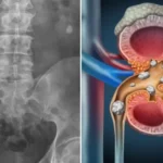
- Imaging Tests: Non-contrast CT scans are the gold standard. Ultrasound may be used in pregnant patients or children.
- Urinalysis: Detects blood, crystals, and signs of infection.
- Blood Tests: Assesses kidney function and identifies metabolic abnormalities.
- 24-Hour Urine Analysis: Measures levels of calcium, oxalate, citrate, and other compounds.
Management and Treatment
Managing calcium oxalate renal calculi involves a combination of dietary changes, medical therapies, and, in some cases, surgical interventions.
1. Dietary Modifications
- Increase Fluid Intake: Aim for at least 2.5-3 liters of water daily to dilute urine.
- Moderate Oxalate Consumption: Limit high-oxalate foods while maintaining a balanced diet.
- Reduce Sodium: Lowering salt intake reduces urinary calcium excretion.
- Adequate Calcium Intake: Dietary calcium binds to oxalate in the gut, reducing its absorption.
2. Medications
- Thiazide Diuretics: Reduce urinary calcium excretion.
- Potassium Citrate: Increases citrate levels to inhibit crystal aggregation.
- Allopurinol: Prescribed for patients with hyperuricosuria.
3. Surgical Interventions
- Extracorporeal Shock Wave Lithotripsy (ESWL): Uses shock waves to break stones into smaller fragments.
- Ureteroscopy: Involves using a thin scope to remove or break up stones.
- Percutaneous Nephrolithotomy (PCNL): Reserved for larger or complex stones.
Prevention
Preventive strategies are key to reducing the recurrence of calcium oxalate stones:
- Stay hydrated consistently.
- Follow a balanced diet low in sodium and oxalate.
- Monitor and address underlying medical conditions.
- Regular follow-ups with a healthcare provider.

