Peripheral edema refers to the accumulation of excess fluid in the interstitial tissues, most commonly observed in the lower extremities such as the feet, ankles, and legs. It is not a disease in itself but rather a symptom indicative of underlying systemic or localized conditions. The pathophysiology involves an imbalance between capillary filtration and lymphatic drainage, leading to visible swelling.
Persistent or recurrent peripheral edema necessitates a thorough clinical assessment to determine its etiology and implement appropriate therapeutic strategies.
Pathophysiology of Edema Formation
Peripheral edema arises from various disturbances in hydrostatic pressure, oncotic pressure, capillary permeability, or lymphatic obstruction. These mechanisms include:
- Increased hydrostatic pressure (e.g., heart failure, venous insufficiency)
- Reduced plasma oncotic pressure (e.g., hypoalbuminemia from liver or kidney disease)
- Capillary permeability elevation (e.g., inflammation, allergic reactions)
- Lymphatic obstruction (e.g., lymphedema, malignancy)
Common Causes of Peripheral Edema
Cardiovascular Disorders
- Congestive Heart Failure (CHF): Right-sided heart failure leads to systemic venous congestion, causing bilateral leg swelling.
- Deep Vein Thrombosis (DVT): A clot obstructs venous return, typically resulting in unilateral edema.
Renal Disorders
- Nephrotic Syndrome: Proteinuria causes hypoalbuminemia, decreasing oncotic pressure and resulting in generalized edema.
- Acute Kidney Injury or Chronic Kidney Disease: Impaired fluid excretion contributes to systemic fluid overload.
Hepatic Conditions
- Cirrhosis: Portal hypertension and reduced albumin synthesis lead to ascites and peripheral swelling.
Endocrine and Metabolic Factors
- Hypothyroidism: Myxedema can manifest as non-pitting edema.
- Diabetes Mellitus: Often leads to vascular damage and fluid imbalance.
Medications
- Calcium channel blockers (e.g., amlodipine)
- NSAIDs
- Steroids
- Hormonal therapies
These agents may alter capillary dynamics or renal function.
Lymphatic and Venous Disorders
- Chronic venous insufficiency: Damaged venous valves cause retrograde flow and pooling.
- Lymphedema: Obstruction or congenital defects in lymphatic drainage result in non-pitting, often unilateral edema.
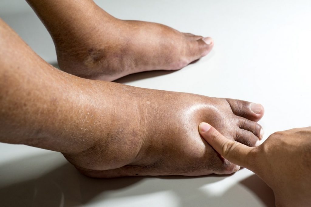
Clinical Classification: Pitting vs. Non-Pitting Edema
- Pitting Edema: Characterized by a lasting depression when pressure is applied to the swollen area. Typically caused by fluid overload or hypoalbuminemia.
- Non-Pitting Edema: Associated with lymphatic obstruction or myxedema. The skin feels firm and thickened without indentation.
Symptoms and Physical Examination Findings
Peripheral edema is often accompanied by:
- Swelling of feet, ankles, or legs
- Tightness or heaviness in limbs
- Skin changes such as shiny or stretched appearance
- Reduced mobility
- Pain or discomfort, especially in cases of DVT or cellulitis
Bilateral edema generally indicates systemic conditions, while unilateral swelling suggests localized pathology.
Diagnostic Approach
A structured diagnostic approach ensures appropriate identification of underlying causes.
Laboratory Investigations
- Complete blood count (CBC) – Detects infection or anemia.
- Serum albumin and liver function tests – Evaluate hepatic synthesis.
- Renal function tests – Assess BUN, creatinine, and urinalysis for proteinuria.
- Thyroid profile – Confirms hypothyroidism.
- D-dimer – Screens for thrombosis if DVT is suspected.
Imaging Studies
- Doppler ultrasound – Essential to detect DVT or venous insufficiency.
- Echocardiography – Assesses cardiac output and right ventricular function.
- CT or MRI – May be indicated in malignancy or lymphatic obstruction.
- Lymphoscintigraphy – Visualizes lymphatic flow in suspected lymphedema.
Evidence-Based Treatment of Peripheral Edema
1. Treating the Underlying Cause
Management depends on addressing the precipitating condition:
- Heart failure – Use of diuretics, ACE inhibitors, beta-blockers
- Renal disease – Sodium restriction, dialysis if needed
- Cirrhosis – Diuretics, paracentesis, TIPS in refractory cases
- Hypothyroidism – Thyroid hormone replacement
- Infection – Antibiotic therapy for cellulitis or abscess
2. Pharmacologic Therapy
- Loop diuretics (furosemide, bumetanide) – Mainstay for fluid removal
- Thiazide diuretics – Used in mild to moderate cases
- Aldosterone antagonists (spironolactone) – Beneficial in cirrhosis and heart failure
Monitoring for electrolyte imbalances is critical during diuretic therapy.
3. Compression Therapy
- Graduated compression stockings improve venous return.
- Manual lymphatic drainage and pneumatic pumps for lymphedema.
4. Lifestyle and Supportive Measures
- Salt restriction (<2 g/day)
- Leg elevation above heart level
- Regular physical activity to enhance circulation
- Weight loss to reduce venous pressure
Complications of Untreated Peripheral Edema
Chronic or severe edema can lead to:
- Skin ulcerations and infections
- Venous stasis dermatitis
- Lymphorrhea and tissue breakdown
- Functional impairment and mobility restriction
Timely diagnosis and management are essential to prevent these sequelae.
Peripheral edema is a multifactorial condition requiring a holistic diagnostic and therapeutic approach. By identifying the underlying etiology—whether cardiovascular, renal, hepatic, endocrine, or iatrogenic—we can implement targeted treatments that alleviate symptoms and prevent recurrence. Long-term management, including lifestyle changes, pharmacotherapy, and monitoring, is essential to ensure optimal vascular and systemic health.