Pemphigus encompasses a group of rare, chronic autoimmune blistering diseases affecting the skin and mucous membranes. It is characterized by the production of autoantibodies directed against desmosomal proteins, essential for keratinocyte adhesion. The resulting acantholysis leads to intraepidermal blister formation, erosions, and significant morbidity if untreated.
Types of Pemphigus: Clinical Variants and Their Distinctions
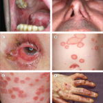
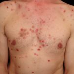
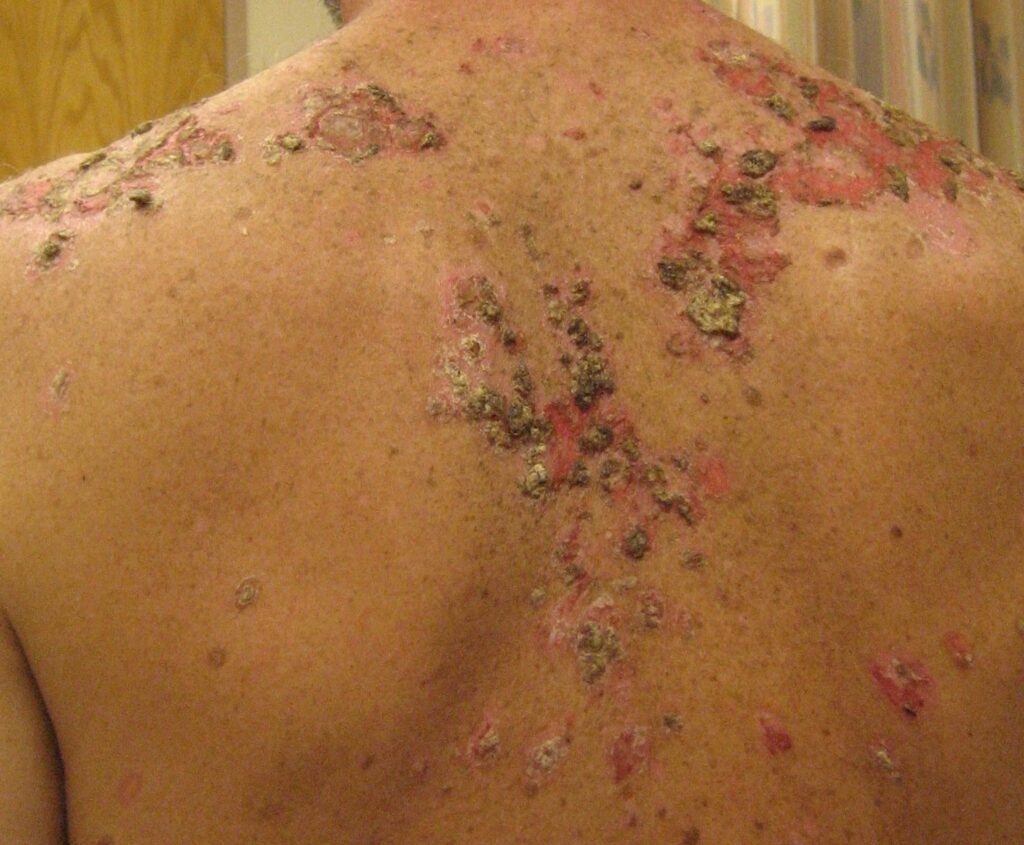
Pemphigus Vulgaris (PV)
Pemphigus vulgaris is the most prevalent and severe form. It typically presents between the third and sixth decades of life. Clinical manifestations begin with painful mucosal erosions, especially in the oral cavity, followed by flaccid skin blisters that rupture easily, leaving widespread denuded areas.
- Target antigen: Desmoglein 3 (mucosal) and Desmoglein 1 (mucocutaneous)
- Commonly involves scalp, face, chest, axillae, and groin
Pemphigus Foliaceus (PF)
This superficial variant primarily involves the skin and spares the mucous membranes. Patients exhibit scaly, crusted lesions often mistaken for seborrheic dermatitis.
- Target antigen: Desmoglein 1
- Commonly affects face, scalp, upper chest, and back
Paraneoplastic Pemphigus (PNP)
Associated with underlying neoplasms, particularly lymphoproliferative disorders. PNP is the most aggressive form, involving severe mucosal erosions, polymorphic skin lesions, and systemic involvement.
- Frequently linked to non-Hodgkin lymphoma, chronic lymphocytic leukemia, and Castleman disease
IgA Pemphigus
Characterized by IgA autoantibodies targeting desmosomal components. This rare form features pustular lesions, often arranged in annular or circinate patterns.
Pathophysiology of Pemphigus: Mechanism of Autoimmune Skin Disruption
Pemphigus results from a Type II hypersensitivity reaction, where autoantibodies, primarily IgG, target desmoglein proteins (cadherins within desmosomes) responsible for intercellular adhesion in the epidermis.
Immunologic Cascade
- Autoantibody production against desmogleins
- Disruption of desmosomal adhesion
- Acantholysis – loss of cohesion between keratinocytes
- Intraepidermal blister formation
- Secondary infection and fluid loss risks
Clinical Manifestations: Recognizing Pemphigus Symptoms
Common Signs and Symptoms
- Painful oral ulcers (initial presentation in PV)
- Flaccid bullae that rupture easily, leaving raw erosions
- Nikolsky’s sign: gentle pressure on skin causes sloughing
- Positive Asboe-Hansen sign: lateral extension of blister with pressure
- Crusting and oozing skin lesions in PF
- Conjunctival and genital involvement in severe cases
Diagnostic Approach: Confirming a Pemphigus Diagnosis
Clinical Evaluation
Diagnosis begins with clinical suspicion, especially in patients presenting with mucocutaneous erosions and flaccid blisters.
Histopathology
- Biopsy reveals intraepidermal acantholysis
- Tzanck smear shows acantholytic (rounded) keratinocytes
Direct Immunofluorescence (DIF)
- Perilesional biopsy demonstrates intercellular IgG and C3 deposition in a “chicken wire” pattern across the epidermis.
Indirect Immunofluorescence (IIF)
- Identifies circulating anti-desmoglein autoantibodies in patient serum.
ELISA
- Quantifies anti-desmoglein 1 and 3 IgG antibodies, aiding in disease severity assessment and monitoring treatment response.
Differential Diagnosis
- Bullous pemphigoid (subepidermal blistering)
- Erythema multiforme
- Stevens-Johnson syndrome
- Herpetic stomatitis
- Lichen planus
Accurate differentiation is crucial due to differences in pathogenesis and management.
Pemphigus Treatment: Modern Therapeutic Strategies
Systemic Corticosteroids
Prednisone remains the cornerstone of therapy for moderate to severe disease. High-dose regimens are employed initially and tapered based on response.
Steroid-Sparing Immunosuppressants
- Azathioprine
- Mycophenolate mofetil
- Cyclophosphamide
- Methotrexate
Used in combination with corticosteroids to reduce dosage and long-term side effects.
Biologic Therapies
- Rituximab: Anti-CD20 monoclonal antibody; highly effective in achieving remission and reducing relapses.
- IVIG (Intravenous Immunoglobulin): Employed in severe or recalcitrant cases.
Adjunctive Therapies
- Plasmapheresis or immunoadsorption to remove circulating autoantibodies
- Dapsone for IgA pemphigus
- Topical corticosteroids for limited disease
Disease Monitoring and Follow-Up
- Regular monitoring of anti-desmoglein antibody titers
- Frequent assessment for treatment-related side effects (osteoporosis, hyperglycemia, infections)
- Multidisciplinary care involving dermatologists, dentists, ophthalmologists, and nutritionists
Prognosis and Complications
With modern immunosuppressive therapy, the mortality rate has significantly decreased, but relapses are common. Untreated pemphigus can lead to:
- Sepsis
- Electrolyte imbalances
- Malnutrition
- Psychological distress due to disfigurement
Early diagnosis and comprehensive treatment are vital for improving long-term outcomes.
Preventive Measures and Patient Education
- Avoidance of known drug triggers (e.g., penicillamine, captopril)
- Prompt reporting of new erosions or infections
- Adherence to treatment regimen
- Nutritional support for mucosal involvement
- Dental care for oral pemphigus
- Psychological counseling for chronic illness support
Pemphigus represents a complex autoimmune dermatological disorder with a potentially devastating impact if not managed promptly. Accurate diagnosis, aggressive immunosuppressive therapy, and vigilant follow-up are key to disease control and improving quality of life. Advancements in biologic therapies such as rituximab have transformed the treatment paradigm, offering hope for sustained remission and recovery.