Parathyroid hyperactivity, typically associated with primary hyperparathyroidism, results from excessive secretion of parathyroid hormone (PTH). Accurate localization of hyperfunctioning parathyroid tissue is essential to guide surgical intervention, particularly for patients undergoing minimally invasive parathyroidectomy. The success of treatment depends heavily on precise preoperative and intraoperative identification of the overactive gland(s).
Pathophysiology of Parathyroid Hyperactivity
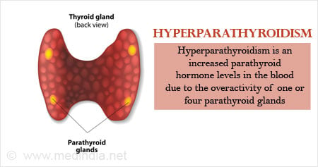
The condition arises from either a solitary adenoma, parathyroid hyperplasia, multiple adenomas, or, rarely, carcinoma. Elevated PTH levels increase serum calcium through enhanced bone resorption, renal calcium reabsorption, and increased intestinal absorption via activated vitamin D.
Indications for Localization in Hyperparathyroidism
Localization studies are particularly indicated for:
- Confirmed biochemical diagnosis of primary hyperparathyroidism
- Minimally invasive parathyroidectomy (MIP) planning
- Reoperative surgery for persistent or recurrent disease
- Ectopic parathyroid adenomas
- Multiglandular disease differentiation
Intraoperative localization techniques may also be employed during surgery when preoperative imaging is inconclusive.
Preoperative Localization Imaging Modalities
1. 99mTc-Sestamibi Scan
- Mechanism: Uses technetium-99m-labeled sestamibi, which preferentially accumulates in mitochondria-rich adenomas.
- Sensitivity: ~80–90% for solitary adenomas.
- Protocol: Dual-phase planar imaging or SPECT/SPECT-CT for 3D localization.
- Advantages: Widely available, non-invasive, effective in solitary lesions.
- Limitations: Reduced accuracy in multiglandular disease and hyperplasia.
2. Ultrasound (USG) of the Neck
- High-resolution neck ultrasound complements sestamibi scans.
- Operator-dependent, best for glands >1 cm.
- Useful in identifying coexisting thyroid nodules and evaluating cervical lymph nodes.
- Limited in retrosternal or ectopic parathyroid localization.
3. 4D-Computed Tomography (4D-CT)
- Combines three-dimensional imaging with temporal perfusion phases (pre-contrast, arterial, and delayed phases).
- Identifies adenomas based on rapid contrast uptake and washout.
- High sensitivity, especially in reoperative settings and ectopic glands.
- Radiation exposure is higher compared to ultrasound or sestamibi.
4. Magnetic Resonance Imaging (MRI)
- Preferred in patients with contraindications to iodinated contrast or radiation.
- Moderate sensitivity but useful in specific anatomical regions like the mediastinum.
- Functional MRI techniques (e.g., dynamic contrast-enhanced MRI) are being evaluated for improved detection.
5. Positron Emission Tomography (PET)
- 18F-fluorocholine PET/CT has emerged as a promising modality.
- High sensitivity in detecting parathyroid adenomas, particularly in cases where conventional imaging fails.
- Useful in persistent or recurrent hyperparathyroidism.
Comparative Overview of Localization Modalities
| Imaging Technique | Sensitivity | Usefulness | Limitations |
|---|---|---|---|
| Sestamibi Scan | 80–90% | Solitary adenoma | Poor for hyperplasia |
| Neck Ultrasound | 70–80% | Surface glands, thyroid eval | Operator-dependent |
| 4D-CT | >90% | Ectopic glands, reoperative cases | High radiation |
| MRI | ~70% | Mediastinal glands | Lower resolution |
| 18F-Choline PET | >95% | Recurrent/persistent disease | Limited availability |
Intraoperative Localization Techniques
1. Intraoperative PTH Monitoring
- Rapid intraoperative assay of serum PTH before and after excision.
- A drop of >50% within 10 minutes confirms removal of hyperfunctioning tissue.
- Essential in multiglandular or ambiguous imaging cases.
2. Radioguided Parathyroidectomy
- Utilizes gamma probe after preoperative sestamibi injection.
- Helps localize ectopic glands and confirm removal intraoperatively.
3. Frozen Section Histology
- Confirms parathyroid tissue intraoperatively but does not distinguish between adenoma and hyperplasia with certainty.
Ectopic and Multiglandular Parathyroid Localization
Ectopic Locations
- Intrathymic, mediastinal, intrathyroidal, retroesophageal, and carotid sheath
- Frequently missed by conventional imaging
- 4D-CT and PET/CT are preferred modalities
Multiglandular Disease
- Accounts for ~10–15% of cases
- Requires careful biochemical correlation and multiple imaging studies
- May necessitate bilateral neck exploration
Surgical Planning Based on Localization
Minimally Invasive Parathyroidectomy (MIP)
- Preferred for patients with positive and concordant localization studies
- Targeted removal of adenoma through a small incision
- Reduces operative time, hospital stay, and complications
Bilateral Neck Exploration
- Indicated in non-localizing or discordant imaging
- Standard approach for suspected multiglandular disease
Reoperative Parathyroid Surgery
- Higher risk due to scar tissue and altered anatomy
- Requires advanced imaging (4D-CT, PET/CT) and intraoperative guidance
- High-volume centers recommended for optimal outcomes
Current Challenges and Evolving Technologies
- False negatives in multiglandular disease remain a concern
- Limited resolution for deep or ectopic glands
- Emerging use of artificial intelligence (AI) and machine learning in imaging analysis
- Hybrid imaging techniques (e.g., PET/MRI) under clinical investigation
Frequently Asked Questions:
What is the best scan to localize parathyroid adenomas?
A combination of sestamibi scan and neck ultrasound is commonly used. 4D-CT or 18F-choline PET/CT may be used when initial imaging is inconclusive.
Can parathyroid hyperplasia be localized?
Localization of hyperplasia is challenging. Imaging may be less sensitive, often necessitating bilateral exploration and intraoperative PTH monitoring.
Is localization always necessary before surgery?
Yes, especially for minimally invasive surgery. Accurate localization reduces operative time and improves surgical success rates.
What if imaging studies are negative?
Negative imaging does not exclude disease. In such cases, bilateral neck exploration and intraoperative monitoring are necessary.
How accurate is 4D-CT for parathyroid localization?
4D-CT has a sensitivity exceeding 90%, particularly in complex or reoperative scenarios, and is highly effective for ectopic glands.
Effective localization of hyperfunctioning parathyroid tissue is critical in the management of parathyroid hyperactivity. With advances in imaging modalities like 4D-CT and 18F-choline PET, surgeons can now achieve high localization accuracy, enabling targeted, minimally invasive procedures. Optimal outcomes require a multidisciplinary approach, leveraging both preoperative imaging and intraoperative adjuncts for precise gland identification.