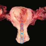
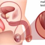
Malignant perivascular epithelioid cell tumors (PEComas) are a rare group of mesenchymal neoplasms characterized by the presence of distinctive perivascular epithelioid cells. These tumors can arise in various body locations, including the uterus, lungs, kidneys, and gastrointestinal tract. While PEComas are often benign, some exhibit malignant behavior, leading to aggressive progression and metastasis.
Etiology and Pathogenesis
The precise cause of PEComas remains unclear; however, genetic and molecular alterations play a crucial role in their development. Notably, a significant number of PEComas are associated with mutations in the TSC1 and TSC2 genes, leading to dysregulation of the mTOR (mechanistic target of rapamycin) pathway. This pathway controls cell growth and proliferation, and its dysregulation contributes to tumorigenesis.
Clinical Presentation
Common Symptoms:
- Abdominal or pelvic mass (if located in the gastrointestinal tract or uterus)
- Pain or discomfort in affected areas
- Hematuria (if renal involvement is present)
- Respiratory issues (if lung involvement is present)
- Weight loss and fatigue in advanced cases
Diagnosis
Imaging Studies
- Ultrasound: Initial evaluation for abdominal or pelvic tumors
- Computed Tomography (CT) Scan: Provides detailed tumor localization and extent
- Magnetic Resonance Imaging (MRI): Useful for soft tissue differentiation
- Positron Emission Tomography (PET) Scan: Detects metastasis and tumor activity
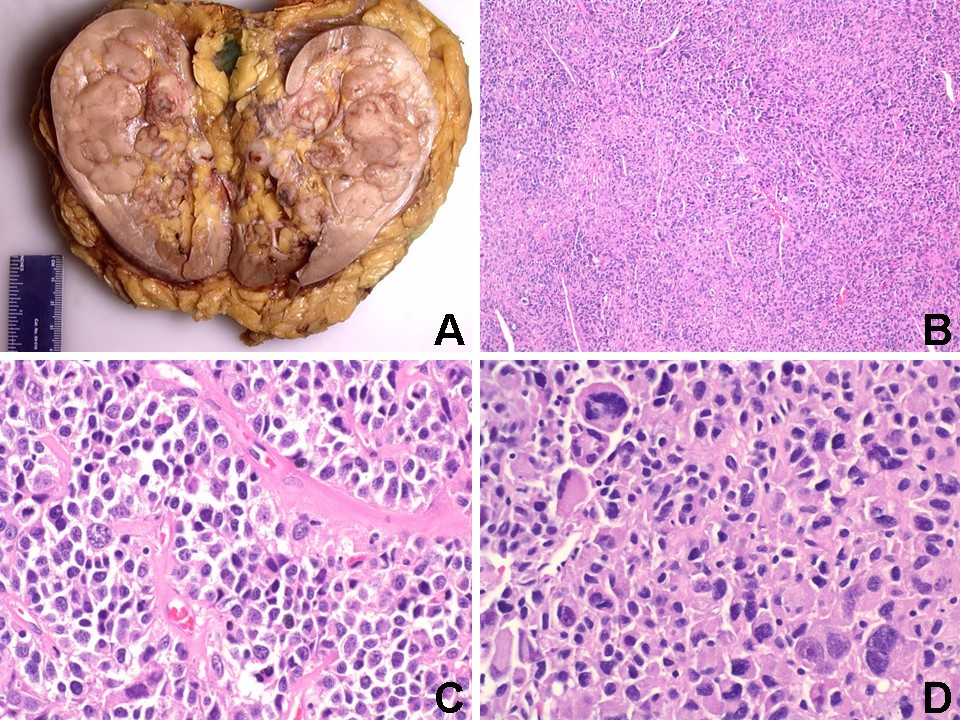
Histopathological Examination
A definitive diagnosis requires a biopsy followed by histopathological analysis, which identifies:
- Large, epithelioid cells with clear to eosinophilic cytoplasm
- Perivascular distribution of tumor cells
- Expression of melanocytic (HMB-45, Melan-A) and muscle markers (SMA, Desmin)
Treatment Approaches
1. Surgical Resection
Complete surgical removal remains the gold standard for localized PEComas. Wide excision with clear margins is crucial to prevent recurrence.
2. Targeted Therapy (mTOR Inhibitors)
Since many PEComas exhibit mTOR pathway activation, inhibitors like sirolimus and everolimus have shown promising results in controlling tumor progression.
3. Chemotherapy
In cases where surgery is not an option or in advanced stages, doxorubicin-based regimens and gemcitabine plus docetaxel have been utilized with limited success.
4. Radiation Therapy
Though not a primary treatment, radiation therapy may be considered for inoperable or metastatic PEComas to manage symptoms and slow tumor growth.
Prognosis and Survival Rates
The prognosis of malignant PEComas depends on various factors, including:
- Tumor size and location
- Histological features (high mitotic index and necrosis indicate worse prognosis)
- Extent of metastasis (lungs and liver are common metastatic sites)
Patients with localized, completely resected tumors have a better prognosis compared to those with metastatic or recurrent disease.
Malignant PEComa is a rare and challenging neoplasm with variable clinical outcomes. Early detection, comprehensive histopathological analysis, and advances in targeted therapies, particularly mTOR inhibitors, offer hope for improved management. Ongoing research continues to refine therapeutic strategies, enhancing survival and quality of life for affected patients.