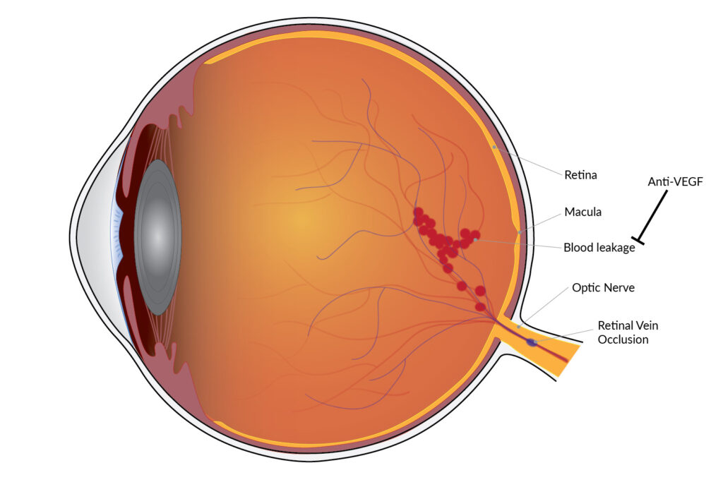
Macular edema following retinal vein occlusion (RVO) is a leading cause of vision impairment, affecting millions worldwide. It occurs when fluid accumulates in the macula due to increased vascular permeability, leading to vision distortion or loss. Understanding the underlying causes, symptoms, and treatment options is essential for effective management and improved patient outcomes.

What is Retinal Vein Occlusion?
Retinal vein occlusion (RVO) is a blockage in the retinal veins, leading to increased pressure, hemorrhages, and leakage of fluids into the macula. It is categorized into two main types:
- Branch Retinal Vein Occlusion (BRVO): Partial blockage affecting a segment of the retina.
- Central Retinal Vein Occlusion (CRVO): Complete blockage of the central retinal vein affecting the entire retina.
How Does RVO Cause Macular Edema?
When the retinal vein is occluded, blood flow is disrupted, causing fluid and protein leakage into the macula. The accumulation of this fluid leads to swelling (edema), distorting central vision. If left untreated, it can result in permanent visual impairment.
Risk Factors for Macular Edema in RVO
Several conditions and lifestyle factors contribute to the development of macular edema following RVO:
- Hypertension – High blood pressure damages blood vessels, increasing the risk of occlusion.
- Diabetes Mellitus – Poor blood sugar control leads to vascular damage and inflammation.
- Hyperlipidemia – High cholesterol contributes to plaque formation, leading to vascular blockages.
- Glaucoma – Increased intraocular pressure affects retinal blood flow.
- Age and Genetics – Older adults and those with a family history of vascular diseases are more susceptible.
Symptoms of Macular Edema After RVO
Patients with macular edema following RVO often experience:
- Blurred or distorted central vision
- Difficulty reading or recognizing faces
- Dark spots or floaters in vision
- Sensitivity to light
- Gradual or sudden vision loss
Diagnosis of Macular Edema in RVO
Early diagnosis is crucial for effective treatment. Eye specialists use several diagnostic techniques:
Optical Coherence Tomography (OCT)
OCT provides high-resolution cross-sectional images of the retina, allowing precise measurement of macular thickness and fluid accumulation.
Fluorescein Angiography (FA)
FA uses a dye injected into the bloodstream to highlight retinal blood vessels, identifying areas of leakage and non-perfusion.
Fundus Photography
This imaging technique captures detailed images of the retina, revealing hemorrhages, swelling, and vascular abnormalities.
Treatment Options for Macular Edema Following RVO
Anti-VEGF Injections
Anti-vascular endothelial growth factor (VEGF) drugs such as ranibizumab (Lucentis) and aflibercept (Eylea) help reduce abnormal blood vessel growth and decrease fluid leakage, improving vision over time.
Corticosteroid Therapy
Intravitreal corticosteroid injections, like dexamethasone implants (Ozurdex), reduce inflammation and fluid retention. However, they carry a risk of increasing intraocular pressure.
Laser Photocoagulation
Focal/grid laser therapy seals leaking blood vessels, reducing fluid accumulation in the macula. While not a primary treatment, it is beneficial in specific cases.
Systemic Management
Controlling underlying conditions such as hypertension, diabetes, and hyperlipidemia is essential for preventing recurrent occlusions and further vision loss.
Prognosis and Long-Term Management
The prognosis varies based on the severity of RVO and the effectiveness of treatment. Regular follow-ups, lifestyle modifications, and adherence to prescribed therapies play a vital role in preserving vision.
Macular edema following retinal vein occlusion is a serious condition requiring timely diagnosis and treatment. With advancements in anti-VEGF therapy, laser treatment, and systemic disease control, patients can achieve significant vision improvement. Consulting an eye specialist at the earliest sign of symptoms ensures better outcomes and long-term eye health.