Gastrointestinal (GI) radiography is pivotal in diagnosing various GI tract disorders. To improve diagnostic accuracy and patient outcomes, adjunct techniques are employed alongside standard imaging methods. This article delves into these adjunct techniques, elucidating their applications and benefits in GI radiography.
Contrast Agents in GI Radiography
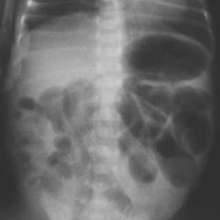
Contrast agents are substances introduced into the body to enhance the visibility of internal structures during radiographic examinations. In GI radiography, barium sulfate is commonly used due to its radiopaque properties, allowing for clear delineation of the GI tract. These agents are integral in various procedures:
- Upper GI Series: Also known as a barium swallow, this series involves the patient ingesting barium to visualize the pharynx, esophagus, stomach, and duodenum. It aids in detecting abnormalities such as strictures, ulcers, and tumors.
- Lower GI Series: Commonly referred to as a barium enema, this procedure introduces barium into the colon via the rectum to assess the large intestine. It is effective in identifying diverticula, polyps, and colorectal cancers.
Double-Contrast Techniques
Double-contrast techniques involve the use of both a radiopaque contrast medium (like barium) and a gas (such as air) to provide a more detailed view of the GI mucosa. The introduction of air distends the GI tract, allowing for better visualization of the mucosal surface and detection of subtle lesions. This method is particularly useful in:
- Double-Contrast Barium Enema: Enhances the detection of small polyps and early mucosal changes in the colon, improving the sensitivity of the examination.
Fluoroscopy in GI Radiography
Fluoroscopy provides real-time X-ray imaging, enabling dynamic assessment of the GI tract. This technique is invaluable for evaluating functional aspects such as swallowing, peristalsis, and sphincter activity. Applications include:
- Barium Swallow Studies: Assess esophageal motility disorders, strictures, and achalasia.
- Small Bowel Follow-Through: Monitors the passage of contrast through the small intestine to detect obstructions or inflammatory diseases.
Cross-Sectional Imaging Adjuncts
While traditional radiography offers valuable information, integrating cross-sectional imaging modalities can enhance diagnostic capabilities:
- Computed Tomography (CT): Provides detailed cross-sectional images, useful in assessing complications like perforations or abscesses.
- Magnetic Resonance Imaging (MRI): Offers superior soft tissue contrast without radiation exposure, beneficial in evaluating fistulas or tumors.
Safety and Efficacy Considerations
The application of adjunct techniques in GI radiography should balance diagnostic benefits with patient safety:
- Radiation Exposure: Minimize exposure by adhering to the ALARA (As Low As Reasonably Achievable) principle.
- Allergic Reactions: Screen patients for allergies to contrast agents to prevent adverse reactions.
- Patient Comfort: Employ measures to reduce discomfort during procedures, such as using appropriate sedation when necessary.
Adjunct techniques in gastrointestinal radiography significantly enhance the detection and characterization of GI disorders. Utilizing contrast agents, double-contrast methods, fluoroscopy, and cross-sectional imaging modalities contributes to more accurate diagnoses and improved patient management. Continuous advancements in these adjunct techniques promise further improvements in GI radiographic practices.