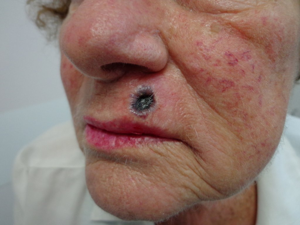
Ecthyma gangrenosum (EG) is a rare but serious cutaneous infection, primarily associated with Pseudomonas aeruginosa bacteremia. First described in 1897 by Dr. Lewellys Barker, EG manifests as necrotic skin ulcers with characteristic erythematous borders. While P. aeruginosa remains the most common causative agent, other pathogens have also been implicated. This condition predominantly affects immunocompromised individuals and necessitates prompt medical intervention.

Etiology
The primary pathogen associated with EG is Pseudomonas aeruginosa, a gram-negative, rod-shaped bacterium commonly found in moist environments. However, EG-like lesions have also been linked to other microorganisms, including:
- Gram-positive bacteria: Staphylococcus aureus, Streptococcus pyogenes
- Gram-negative bacteria: Escherichia coli, Klebsiella pneumoniae, Citrobacter freundii
- Fungi: Candida species, Aspergillus fumigatus
- Viruses: Herpes simplex virus
Immunocompromised states, such as neutropenia, leukemia, diabetes mellitus, malnutrition, and extensive burn wounds, increase susceptibility to EG. Notably, while EG predominantly affects immunocompromised individuals, cases have been reported in immunocompetent persons as well.
Pathophysiology
EG can develop through two primary mechanisms:
- Bacteremic Route: Hematogenous dissemination of the pathogen leads to perivascular invasion, causing ischemic necrosis of the skin.
- Non-bacteremic Route: Direct inoculation of the pathogen into the skin results in localized infection.
Virulence factors produced by P. aeruginosa, such as exotoxin A, elastase, and phospholipase C, contribute to tissue destruction and necrosis.
Clinical Features
EG lesions typically progress through the following stages:
- Initial Stage: Painless, round, erythematous macules or patches.
- Progressive Stage: Development of central pustules with surrounding erythema.
- Advanced Stage: Formation of hemorrhagic vesicles that evolve into necrotic ulcers with black eschars and erythematous halos.
These lesions can develop rapidly, transitioning to necrotic ulcers within 12 to 24 hours. Commonly affected areas include the anogenital region, axillae, extremities, trunk, and face.
Diagnosis
Accurate diagnosis of EG involves:
- Clinical Examination: Identification of characteristic skin lesions.
- Laboratory Tests:
- Blood Cultures: To detect bacteremia.
- Wound Cultures: To identify the causative organism.
- Skin Biopsy: Histopathological examination revealing necrotic epidermis and dermis with perivascular bacterial invasion.
Utilizing a Wood’s lamp may aid in diagnosis, as P. aeruginosa exhibits green fluorescence under ultraviolet light.
Treatment
Immediate initiation of empiric antibiotic therapy is crucial. Recommended treatments include:
- Antipseudomonal Agents:
- Beta-lactams: Piperacillin/tazobactam
- Cephalosporins: Cefepime
- Fluoroquinolones: Levofloxacin
- Carbapenems: Imipenem
Combination therapy is often advised for high-risk patients, such as those with neutropenia or septic shock. Once culture results and sensitivities are available, antibiotic therapy should be tailored accordingly. In cases involving fungal or viral pathogens, appropriate antifungal or antiviral treatments should be administered.
Surgical intervention, including debridement or incision and drainage, may be necessary for extensive necrotic lesions or abscesses.
Prognosis
The prognosis of EG largely depends on the patient’s underlying health status and the promptness of treatment initiation. Mortality rates range from 20% to 77% in bacteremic cases, particularly among immunocompromised individuals. Non-bacteremic cases have a significantly lower mortality rate, approximately 8%. Neutropenia at the time of diagnosis is a critical prognostic factor.