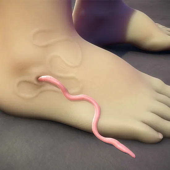
Dracunculiasis, commonly known as Guinea worm disease, is a parasitic infection caused by Dracunculus medinensis. Historically prevalent in rural regions of Africa and Asia, this disease has seen a dramatic decline due to extensive global eradication efforts. As of recent reports, the number of human cases has dwindled to near elimination, but vigilance remains necessary to prevent resurgence.

Lifecycle of Dracunculus medinensis
The lifecycle of D. medinensis is intricate and closely tied to stagnant water sources.
- Ingestion: Humans consume water containing copepods (water fleas) infected with D. medinensis larvae.
- Release and Penetration: In the stomach, copepods are digested, releasing larvae that penetrate the intestinal wall.
- Maturation: Larvae enter the abdominal cavity, maturing into adult worms over 10–14 months.
- Migration: Fertilized female worms migrate to the skin’s surface, typically in the lower limbs.
- Emergence: A blister forms and ruptures, allowing the worm to emerge and release larvae upon contact with water.
- Continuation: Released larvae are ingested by copepods, perpetuating the cycle.
Clinical Manifestations
Approximately one year post-infection, individuals may experience:
- Prodromal Symptoms: Fever, swelling, and pain in the affected area.
- Blister Formation: A painful blister develops, often on the lower limbs, which ruptures to expose the emerging worm.
- Complications: Secondary bacterial infections can lead to abscesses, sepsis, or tetanus.
Diagnosis
Diagnosis is primarily clinical, based on the characteristic appearance of the emerging worm. Radiographic imaging can detect calcified worms from previous infections. Laboratory testing is generally unnecessary due to the disease’s distinct presentation.
Treatment
There are no specific medications to treat dracunculiasis. The traditional method involves:
- Mechanical Extraction: Slowly winding the emerging worm around a stick or gauze, extracting it over days to weeks to prevent breakage.
- Wound Care: Keeping the area clean to prevent secondary infections.
- Pain Management: Administering analgesics to alleviate discomfort.
Prevention and Eradication Efforts
Preventative strategies focus on interrupting transmission:
- Water Filtration: Using fine-mesh filters to remove infected copepods from drinking water.
- Health Education: Informing communities about the importance of consuming filtered or boiled water.
- Case Containment: Encouraging affected individuals to avoid immersing blisters in communal water sources.
- Vector Control: Applying larvicides to water bodies to eliminate copepods.
The global eradication campaign has achieved a reduction in cases from millions in the 1980s to fewer than 20 in recent years. Challenges remain, including infections in animal reservoirs like dogs, which necessitate continued vigilance and adaptive strategies.