Corneal erosion is a condition characterized by the recurrent breakdown of the cornea’s outermost layer, the epithelium. This disruption leads to significant discomfort and potential vision impairment. Understanding the causes, symptoms, and treatment options is crucial for effective management and prevention of complications.
Anatomy of the Cornea
The cornea is the transparent, dome-shaped surface that covers the front of the eye, playing a pivotal role in focusing vision. It comprises five distinct layers:
- Epithelium: The outermost layer, serving as a barrier against dust, bacteria, and other foreign particles.
- Bowman’s Membrane: A tough layer that protects the corneal stroma.
- Stroma: The thick, middle layer composed of collagen fibers, providing structural support.
- Descemet’s Membrane: A thin but strong layer that serves as the basement membrane for the endothelium.
- Endothelium: The innermost layer responsible for maintaining corneal transparency by regulating fluid content.
Causes of Corneal Erosion
Corneal erosion often results from:
- Trauma: Injuries from fingernails, makeup brushes, or tree branches can damage the corneal epithelium.
- Corneal Dystrophies: Genetic conditions like epithelial basement membrane dystrophy (EBMD) weaken epithelial adhesion, increasing susceptibility to erosion.
- Dry Eyes: Insufficient lubrication can lead to epithelial breakdown.
- Previous Corneal Abrasions: Inadequate healing from past abrasions may predispose individuals to recurrent erosions.
Symptoms of Corneal Erosion
Patients with corneal erosion may experience:
- Sudden Eye Pain: Often occurring upon waking, due to the eyelid sticking to the cornea.
- Tearing: Excessive lacrimation as the eye attempts to lubricate the affected area.
- Light Sensitivity (Photophobia): Discomfort in bright environments.
- Blurred Vision: Resulting from irregularities on the corneal surface.
- Foreign Body Sensation: A persistent feeling of something in the eye.
Diagnosis of Corneal Erosion
A comprehensive eye examination is essential for diagnosis:
- Patient History: Assessing symptoms, onset, and any prior eye injuries.
- Slit-Lamp Examination: Utilizing a microscope to inspect the cornea for defects.
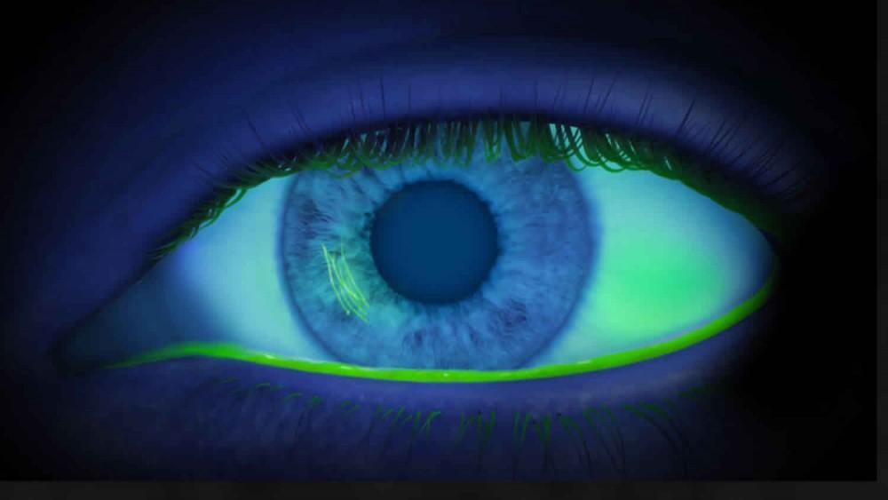
- Fluorescein Staining: Applying a dye to highlight areas of epithelial loss under blue light.
Treatment Options for Corneal Erosion
Management strategies depend on severity and recurrence:
- Medical Management:
- Lubricating Eye Drops or Ointments: To maintain moisture and facilitate healing.
- Antibiotic Drops or Ointments: Preventing secondary bacterial infections.
- Hypertonic Saline Solutions: Reducing corneal edema and promoting epithelial adhesion.
- Bandage Contact Lenses: Protecting the cornea and reducing discomfort during healing.
- Surgical Interventions:
- Debridement: Removing loose or non-adherent epithelium to allow healthy regrowth.
- Anterior Stromal Puncture: Creating tiny punctures in the Bowman’s layer to enhance epithelial attachment.
- Phototherapeutic Keratectomy (PTK): Using a laser to resurface the cornea, beneficial in recurrent or severe cases.
Prevention and Prognosis
Preventive measures include:
- Eye Protection: Wearing safety goggles during activities that pose a risk of eye injury.
- Managing Underlying Conditions: Treating dry eyes or corneal dystrophies appropriately.
- Proper Eyelid Hygiene: Especially in conditions like blepharitis, to reduce inflammation and potential for erosion.
With timely and appropriate treatment, the prognosis for corneal erosion is generally favorable. However, recurrence is possible, necessitating ongoing care and preventive strategies.