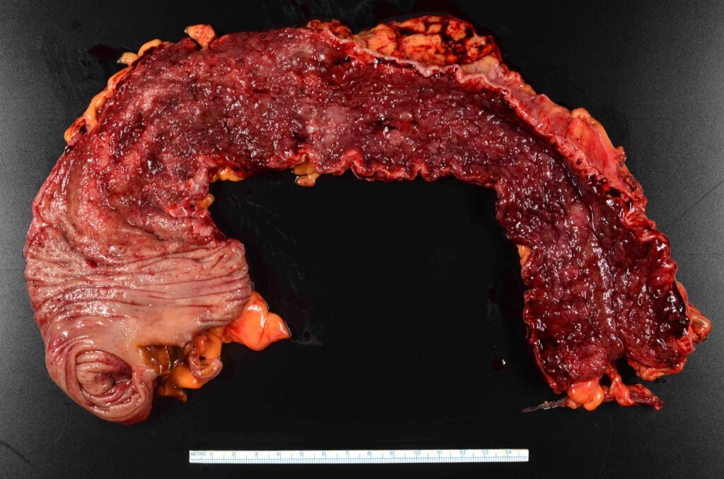
Chronic ulcerative rectosigmoiditis, a subtype of ulcerative colitis, is an inflammatory bowel disease (IBD) characterized by persistent inflammation and ulceration in the rectum and sigmoid colon. This condition impacts the digestive system, leading to various symptoms that can significantly affect a patient’s quality of life. Early diagnosis and effective management are crucial in mitigating complications and improving outcomes.

Symptoms and Clinical Presentation
Patients with chronic ulcerative rectosigmoiditis commonly exhibit the following symptoms:
- Persistent Diarrhea: Often bloody, associated with urgency and tenesmus.
- Abdominal Pain: Cramping in the lower left quadrant, correlating with sigmoid inflammation.
- Rectal Bleeding: Due to mucosal ulceration in the rectum and sigmoid colon.
- Fatigue: Resulting from anemia or chronic inflammation.
- Weight Loss: Occurring in severe or prolonged cases.
- Fecal Incontinence: In advanced or untreated cases.
Complications
- Strictures: Chronic inflammation can lead to scarring and narrowing of the colon.
- Toxic Megacolon: A life-threatening complication requiring immediate medical attention.
- Colorectal Cancer: Increased risk due to prolonged inflammation.
Causes and Risk Factors
Etiology
While the exact cause of chronic ulcerative rectosigmoiditis remains unclear, it is believed to result from an interplay of genetic, immune, and environmental factors.
Risk Factors
- Genetics: Family history of inflammatory bowel disease.
- Immune Dysregulation: Abnormal immune responses targeting the intestinal lining.
- Environmental Triggers: Smoking cessation, infections, or dietary factors.
- Age: Typically diagnosed between 15 and 30 years, but can occur at any age.
- Ethnicity: Higher prevalence in Caucasian populations.
Diagnostic Approach
Clinical Evaluation
A thorough medical history and physical examination are critical for diagnosis.
Diagnostic Tests
- Colonoscopy with Biopsy: The gold standard for diagnosis. Reveals mucosal ulcerations, erythema, and histological inflammation.
- Stool Tests: Rule out infections and detect inflammatory markers such as calprotectin.
- Imaging Studies:
- CT or MRI Enterography: Assess complications like strictures or abscesses.
- Abdominal Ultrasound: Non-invasive evaluation of bowel wall thickening.
Laboratory Investigations
- Complete Blood Count (CBC): Identifies anemia or leukocytosis.
- C-reactive Protein (CRP) and Erythrocyte Sedimentation Rate (ESR): Markers of systemic inflammation.
- Serologic Tests: May reveal autoantibodies like p-ANCA, supporting the diagnosis.
Treatment Strategies
Medical Management
- Aminosalicylates (5-ASA): First-line treatment to reduce inflammation.
- Corticosteroids: For moderate to severe flares, used short-term to control symptoms.
- Immunomodulators:
- Azathioprine or 6-Mercaptopurine (6-MP) for maintenance therapy.
- Biologics like infliximab or adalimumab targeting TNF-α.
- Janus Kinase (JAK) Inhibitors: Emerging options for refractory cases.
- Antibiotics: If secondary infections are suspected.
Surgical Options
Surgical intervention may be required in severe cases or when medical therapy fails. Options include:
- Proctocolectomy with Ileal Pouch-Anal Anastomosis (IPAA): Standard procedure for ulcerative colitis.
- Subtotal Colectomy: For emergency situations such as toxic megacolon.
Lifestyle Modifications and Prevention
Dietary Considerations
- Low-Residue Diet: Reduces bowel irritation during flares.
- Probiotic-Rich Foods: Support gut microbiota balance.
- Hydration: Prevents dehydration from diarrhea.
Stress Management
Chronic stress can exacerbate symptoms. Techniques such as mindfulness, yoga, or cognitive-behavioral therapy may help.
Smoking Cessation
While smoking cessation can initially trigger symptoms, it ultimately benefits long-term health.
Prognosis
With proper management, most patients achieve remission and maintain a good quality of life. However, regular follow-ups are essential to monitor disease progression and prevent complications.