Cervicofacial actinomycosis is a rare chronic bacterial infection primarily caused by Actinomyces species, which are gram-positive, anaerobic bacteria. This condition predominantly affects the head and neck region and is characterized by slowly progressing, suppurative, and granulomatous inflammation. Below, we present a thorough analysis of this condition, including its etiology, clinical manifestations, diagnostic approaches, and treatment options.

Etiology and Pathogenesis
The primary causative agent of cervicofacial actinomycosis is Actinomyces israelii. These bacteria are part of the normal flora of the human oropharynx, gastrointestinal tract, and urogenital tract but can become pathogenic when mucosal barriers are disrupted. Common predisposing factors include:
- Dental procedures (e.g., tooth extractions, root canals)
- Facial trauma
- Poor oral hygiene
- Orofacial surgery
Once the mucosal integrity is compromised, Actinomyces organisms invade surrounding tissues, leading to chronic infection. The hallmark of the disease is the formation of abscesses and sinus tracts that may discharge sulfur granules.
Clinical Features
Cervicofacial actinomycosis typically presents with the following symptoms:
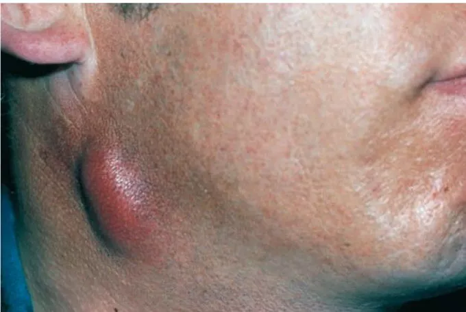
- Swelling and Mass Formation: Painless or mildly tender swelling in the jawline, cheek, or submandibular area.
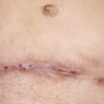
- Sinus Tract Development: Formation of draining sinuses that discharge purulent material containing yellowish sulfur granules.
- Localized Pain: While often mild, pain may increase with secondary bacterial infection.
- Induration and Fibrosis: Chronic cases may lead to firm, woody induration of the affected area.
Commonly affected areas include the submandibular, parotid, and perimandibular regions. Advanced cases may involve contiguous structures, including bones and lymph nodes.
Diagnosis
Accurate diagnosis of cervicofacial actinomycosis relies on clinical suspicion and laboratory confirmation. Key diagnostic steps include:
- Clinical Examination: Noting the characteristic swelling, sinus tracts, and discharge of sulfur granules.
- Microscopic Examination: Identifying gram-positive branching filaments in pus or tissue samples.
- Culture Studies: Isolating Actinomyces species using anaerobic culture techniques.
- Imaging Studies:
- CT or MRI: To evaluate the extent of soft tissue and bony involvement.
- Ultrasound: Useful for identifying abscesses and guiding aspiration.
- Histopathology: Confirmatory biopsy showing chronic granulomatous inflammation with actinomycotic colonies.
Differential Diagnosis
Cervicofacial actinomycosis may mimic other conditions, including:
- Tuberculosis
- Fungal infections (e.g., candidiasis, mucormycosis)
- Malignancies (e.g., squamous cell carcinoma)
- Lymphadenitis
- Osteomyelitis
Treatment and Management
The cornerstone of treatment is prolonged antibiotic therapy combined with surgical intervention when necessary. Recommended management includes:
Antibiotic Therapy
- High-dose Penicillin G: The first-line treatment, typically administered intravenously for 4-6 weeks, followed by oral penicillin or amoxicillin for 6-12 months.
- Alternative Antibiotics: For penicillin-allergic patients, doxycycline, clindamycin, or erythromycin may be used.
Surgical Management
- Abscess Drainage: To relieve pressure and reduce bacterial load.
- Debridement: Excision of necrotic tissue and sinus tract exploration.
- Reconstruction: In advanced cases, reconstructive surgery may be required to restore function and aesthetics.
Prevention
Preventive measures focus on maintaining optimal oral health and minimizing risk factors:
- Regular dental check-ups and cleanings.
- Prompt treatment of dental infections.
- Good oral hygiene practices.
- Avoidance of tobacco and other irritants.
Complications
Untreated or inadequately treated cervicofacial actinomycosis can lead to:
- Chronic Disfigurement: Due to fibrosis and scarring.
- Osteomyelitis: Infection spreading to adjacent bones.
- Systemic Spread: Rarely, hematogenous dissemination to the lungs or other organs.

