Tinea capitis is a contagious dermatophytic fungal infection of the scalp and hair shafts, primarily affecting children but occasionally seen in adults. It is caused by various species of Trichophyton and Microsporum and spreads through direct human-to-human, animal-to-human, or fomite transmission.
Clinically, it manifests in various forms ranging from non-inflammatory scaly patches to severe inflammatory lesions known as kerions. Timely diagnosis and appropriate antifungal treatment are crucial to prevent permanent hair loss and complications.
Causes and Risk Factors of Tinea Capitis
Causative Organisms:
- Trichophyton tonsurans – Most common in North America and UK
- Microsporum canis – Associated with pets like cats and dogs
- Trichophyton violaceum – Common in African and Middle Eastern regions
Modes of Transmission:
- Direct contact with infected individuals or animals
- Sharing personal items like combs, hats, towels
- Poor hygiene and overcrowded living conditions
Predisposing Factors:
- Age (most prevalent in children aged 3–14)
- Immunosuppression
- Frequent exposure to pets or farm animals
- Socioeconomic conditions affecting hygiene standards
Clinical Features and Tinea Capitis Symptoms
Tinea capitis presents with a range of dermatological features depending on the degree of inflammation and fungal species involved.
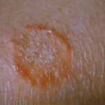
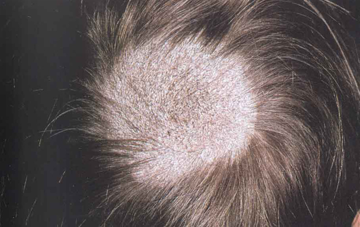
Non-inflammatory (Black Dot & Grey Patch Type):
- Scaly, round patches of hair loss
- Black dots where hairs have broken off at the scalp
- Fine scaling resembling dandruff
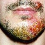
Inflammatory (Kerion and Favus Type):
- Painful, boggy swelling with pus discharge (kerion)
- Crusting, yellow cup-shaped scutula (favus)
- Lymphadenopathy
- Fever and malaise in severe cases
Other Signs:
- Hair fragility and breakage
- Pruritus (itching)
- Secondary bacterial infections
Diagnostic Techniques for Tinea Capitis
Accurate diagnosis is essential for effective treatment and prevention of transmission.
Clinical Assessment:
- Visual inspection of scalp and hair
- Identification of classic signs like black dots, scaling, and alopecia
Laboratory Tests:
- Wood’s Lamp Examination: Microsporum infections fluoresce green under UV light
- Direct Microscopy (KOH Preparation): Shows fungal hyphae in hair shafts
- Fungal Culture: Confirms species-specific dermatophyte
- Dermatoscopy: Identifies comma hairs, corkscrew hairs, and broken shafts
Effective Treatment of Tinea Capitis
Oral antifungal therapy is the cornerstone of treatment due to the deep-seated nature of the infection in the hair shaft.
Systemic Antifungals:
- Griseofulvin (15–25 mg/kg/day for 6–8 weeks): Gold standard in children
- Terbinafine (62.5–250 mg/day based on weight for 4–6 weeks): Effective against Trichophyton species
- Itraconazole or Fluconazole: Alternatives for resistant or complicated cases
Adjunctive Therapy:
- Topical Antifungals: Ketoconazole or selenium sulfide shampoos to reduce spore shedding
- Corticosteroids: In severe inflammatory kerion to reduce swelling and scarring
- Antibiotics: If secondary bacterial infection is present
Prevention of Tinea Capitis and Hygiene Recommendations
Preventing recurrence and limiting transmission requires diligent hygiene practices and public health awareness.
Individual Measures:
- Avoid sharing combs, hats, pillows, and towels
- Regular scalp washing with antifungal shampoos in at-risk environments
- Keep nails trimmed and clean to avoid autoinoculation
Community Strategies:
- Screen and treat infected individuals and close contacts
- Veterinary treatment of infected pets
- School health education programs
- Prompt exclusion of affected children from school until therapy begins
Complications and Long-Term Outcomes
Untreated or inadequately treated tinea capitis can result in significant morbidity.
Potential Complications:
- Permanent scarring alopecia from kerion
- Chronic infection due to poor compliance
- Psychological impact due to cosmetic disfigurement in children
- Secondary bacterial infections
Prognosis:
With early systemic therapy and proper hygiene, the condition resolves without permanent hair loss in most cases. Severe kerion may leave residual scarring.
Key Insights on Tinea Capitis
- Tinea capitis is a contagious scalp fungal infection, most common in children
- Requires systemic antifungal therapy for resolution
- Early identification prevents scarring and spread
- Hygiene and environmental decontamination are key to prevention
Frequently Asked Questions:
What is the most common cause of tinea capitis?
Tinea capitis is most commonly caused by Trichophyton tonsurans, particularly in North America and the UK, and Microsporum canis in pet-associated infections.
Can adults get tinea capitis?
Although rare, adults—especially those who are immunocompromised—can contract tinea capitis, often from infected children or pets.
How is tinea capitis transmitted?
It spreads via direct contact with infected individuals, animals, or contaminated objects like brushes, combs, hats, and bedding.
How long does treatment for tinea capitis take?
Treatment typically lasts between 4 to 8 weeks depending on the antifungal used, severity of infection, and patient compliance.
Can tinea capitis cause permanent hair loss?
Yes, if left untreated—particularly in inflammatory types like kerion—it can result in scarring alopecia and permanent hair loss.