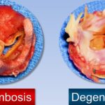
Thromboembolic disorders are conditions characterized by the abnormal formation and migration of blood clots within the circulatory system. These clots, or thrombi, may form in veins or arteries and can travel through the bloodstream, causing embolic events that obstruct blood flow to vital organs. This disruption can lead to severe clinical outcomes, including pulmonary embolism, stroke, myocardial infarction, or even sudden death.
Timely recognition, accurate diagnosis, and effective management are essential for reducing morbidity and mortality associated with thromboembolic events.

Classification of Thromboembolic Disorders
1. Venous Thromboembolism (VTE)
VTE is the most prevalent type of thromboembolic disorder, comprising:
- Deep Vein Thrombosis (DVT): Clot formation in deep veins, typically in the lower extremities.
- Pulmonary Embolism (PE): Occurs when a clot dislodges and travels to the pulmonary arteries, blocking lung circulation.
2. Arterial Thromboembolism
Arterial emboli typically originate in the heart or large arteries and can cause:
- Ischemic Stroke
- Myocardial Infarction (MI)
- Acute Limb Ischemia
- Retinal Artery Occlusion
Pathophysiology of Thromboembolic Disorders
The pathogenesis of thromboembolic disease is rooted in Virchow’s Triad, which outlines three primary factors:
- Endothelial Injury: Vascular trauma, inflammation, or surgery damages the inner lining of blood vessels.
- Hypercoagulability: Genetic or acquired conditions increase the tendency for clot formation (e.g., Factor V Leiden, antiphospholipid syndrome).
- Venous Stasis: Immobility, heart failure, or venous compression reduces blood flow, promoting clot formation.
Risk Factors for Thromboembolic Events
Thromboembolic disorders arise from both genetic predispositions and acquired conditions. Major risk factors include:
- Prolonged immobility or hospitalization
- Surgery, especially orthopedic or abdominal
- Cancer and chemotherapy
- Hormonal therapy and oral contraceptives
- Obesity
- Pregnancy and postpartum period
- Smoking
- Thrombophilia (e.g., Protein C or S deficiency)
- Atrial fibrillation (for arterial thromboembolism)
Signs and Symptoms of Thromboembolic Events
Deep Vein Thrombosis (DVT)
- Unilateral leg swelling
- Pain or tenderness
- Redness and warmth over the vein
Pulmonary Embolism (PE)
- Sudden shortness of breath
- Chest pain, often pleuritic
- Hemoptysis
- Tachycardia or hypotension
- Cyanosis in severe cases
Arterial Embolism
- Sudden pain in affected limb
- Pallor and pulselessness
- Neurologic deficits (if cerebral)
- Coldness and paralysis (limb-threatening ischemia)
Diagnostic Evaluation of Thromboembolic Disorders
Laboratory Tests
- D-dimer: Elevated in acute thrombotic states; useful for excluding DVT/PE in low-risk patients
- Coagulation profile: PT, aPTT, INR
- Thrombophilia panel: In suspected hereditary clotting disorders
Imaging Techniques
- Compression Ultrasonography: First-line for suspected DVT
- CT Pulmonary Angiography (CTPA): Gold standard for PE
- Ventilation-Perfusion (V/Q) Scan: Alternative to CTPA in renal dysfunction or contrast allergy
- Echocardiography: Identifies cardiac sources of embolism
- MRI/MRA: For arterial embolic events, particularly cerebral
Management of Thromboembolic Disorders
1. Anticoagulation Therapy
The cornerstone of thromboembolism treatment is anticoagulation, aimed at halting clot propagation and preventing recurrence.
| Drug Class | Examples | Notes |
|---|---|---|
| Heparins | Unfractionated heparin, Enoxaparin | Used acutely; rapid onset |
| Vitamin K Antagonists | Warfarin | Requires INR monitoring |
| Direct Oral Anticoagulants (DOACs) | Apixaban, Rivaroxaban, Dabigatran | Preferred for long-term outpatient use |
Duration depends on the nature of the event:
- 3–6 months: Provoked thromboembolism
- Indefinite: Unprovoked or recurrent events, or persistent risk factors
2. Thrombolytic Therapy
Indicated in life-threatening PE or massive arterial occlusion when rapid clot dissolution is necessary.
- Alteplase (tPA) is commonly used
- Strict criteria due to bleeding risk
3. Mechanical Interventions
- Inferior Vena Cava (IVC) Filter: For patients contraindicated for anticoagulation
- Surgical or catheter-directed thrombectomy: In severe limb-threatening ischemia or massive PE
4. Management of Underlying Causes
- Cancer therapy
- Cardiac rhythm control in atrial fibrillation
- Discontinuation of prothrombotic medications
Complications of Thromboembolic Disease
- Post-thrombotic syndrome: Chronic leg swelling and pain after DVT
- Pulmonary hypertension: Following recurrent PE
- Stroke and disability: In arterial embolism affecting the brain
- Chronic limb ischemia
Prompt recognition and long-term follow-up are essential to minimize these outcomes.
Prevention Strategies
Primary Prevention
- Early ambulation post-surgery
- Mechanical prophylaxis: Compression stockings, pneumatic devices
- Pharmacologic prophylaxis: Low-dose anticoagulants in high-risk hospitalized patients
Secondary Prevention
- Long-term anticoagulation
- Lifestyle changes: Smoking cessation, weight loss, physical activity
- Management of comorbidities: Hypertension, diabetes, atrial fibrillation
Prognosis and Outlook
With appropriate therapy, the prognosis for thromboembolic disorders can be favorable. However, recurrent events and long-term complications necessitate continued vigilance and management. Patients with unprovoked or cancer-associated thromboembolism have a higher recurrence rate and may require indefinite anticoagulation.
Thromboembolic disorders represent a spectrum of potentially fatal vascular conditions involving pathological clot formation and embolization. Early recognition, individualized anticoagulant management, and mitigation of risk factors are essential to improving patient outcomes. With the rising global burden of thromboembolic disease, particularly in aging and sedentary populations, the focus must remain on early detection, prevention, and comprehensive long-term care.