Surgical procedures on the eye proper refer to interventions involving the ocular globe and its internal structures. These highly specialized operations aim to restore, preserve, or enhance vision by addressing pathological conditions that cannot be treated with pharmacological or non-invasive therapies. Common procedures include cataract extraction, glaucoma filtration surgery, vitrectomy, corneal transplantation, and retinal reattachment, each targeting a specific anatomical component of the eye.

Anatomy of the Eye Proper: A Surgical Perspective
A precise understanding of ocular anatomy is essential for safe and effective surgical planning. The eye proper includes:
- Cornea: Transparent anterior structure essential for refraction.
- Anterior Chamber: Space filled with aqueous humor between the cornea and iris.
- Lens: Biconvex structure responsible for accommodation.
- Vitreous Body: Gel-like substance occupying the posterior segment.
- Retina: Neural layer responsible for phototransduction.
- Choroid and Sclera: Supportive vascular and fibrous layers.
- Optic Nerve: Conveys visual signals to the brain.
Cataract Surgery: Phacoemulsification and Intraocular Lens Implantation
Indications
Cataract surgery is indicated when opacification of the lens leads to significant visual impairment, interfering with daily activities or increasing fall risk.
Technique
- Phacoemulsification: Ultrasonic probe emulsifies the cataractous lens.
- IOL Implantation: A synthetic intraocular lens is inserted through a small corneal incision.
Outcomes
This is the most commonly performed eye surgery worldwide, with high success rates and minimal complication risk when performed by skilled surgeons.
Glaucoma Surgery: Reducing Intraocular Pressure (IOP)
Indications
For patients with progressive optic nerve damage and uncontrolled IOP despite maximal medical therapy.
Surgical Options
- Trabeculectomy: Creates a new drainage pathway.
- Glaucoma Drainage Devices (e.g., Ahmed, Baerveldt): Implanted to shunt aqueous humor externally.
- Minimally Invasive Glaucoma Surgery (MIGS): iStent, Hydrus—used for mild to moderate cases.
Complications
May include hypotony, bleb leaks, or endophthalmitis, requiring careful postoperative monitoring.
Vitrectomy: Posterior Segment Intervention
Indications
Vitrectomy is performed for:
- Retinal detachment
- Macular hole
- Diabetic vitreous hemorrhage
- Epiretinal membrane
Technique
Pars plana vitrectomy involves the removal of the vitreous gel to gain access to the retina. Often combined with membrane peeling, endolaser photocoagulation, or tamponade agents (e.g., gas or silicone oil).
Corneal Surgeries: Restoring Transparency and Curvature
Penetrating Keratoplasty (PK)
Full-thickness corneal transplant used for conditions like keratoconus or corneal scarring.
Deep Anterior Lamellar Keratoplasty (DALK)
Selective replacement of stromal layers, preserving Descemet’s membrane and endothelium.
Descemet Membrane Endothelial Keratoplasty (DMEK)
Selective replacement of the endothelium, preferred for Fuchs’ endothelial dystrophy.
Refractive Surgeries: Vision Correction Beyond Glasses
LASIK (Laser-Assisted In Situ Keratomileusis)
- Reshapes the corneal stroma using excimer laser
- Treats myopia, hyperopia, and astigmatism
PRK (Photorefractive Keratectomy)
- Surface ablation technique without a corneal flap
- Suitable for thinner corneas
SMILE (Small Incision Lenticule Extraction)
- Uses femtosecond laser to extract a stromal lenticule via a microincision
Retinal Surgeries: Addressing Posterior Segment Pathology
Scleral Buckling
Used primarily for rhegmatogenous retinal detachment, the external buckle indents the sclera to close retinal breaks.
Pneumatic Retinopexy
In-office procedure involving intravitreal gas injection followed by laser or cryopexy to seal the retinal tear.
Submacular Surgery
Rarely performed; involves direct access to the macula to remove subretinal neovascular membranes or hemorrhage.
Pediatric and Congenital Eye Surgeries
Congenital Cataract Removal
Performed early in life to prevent amblyopia. Requires careful selection of IOL and often amblyopia therapy postoperatively.
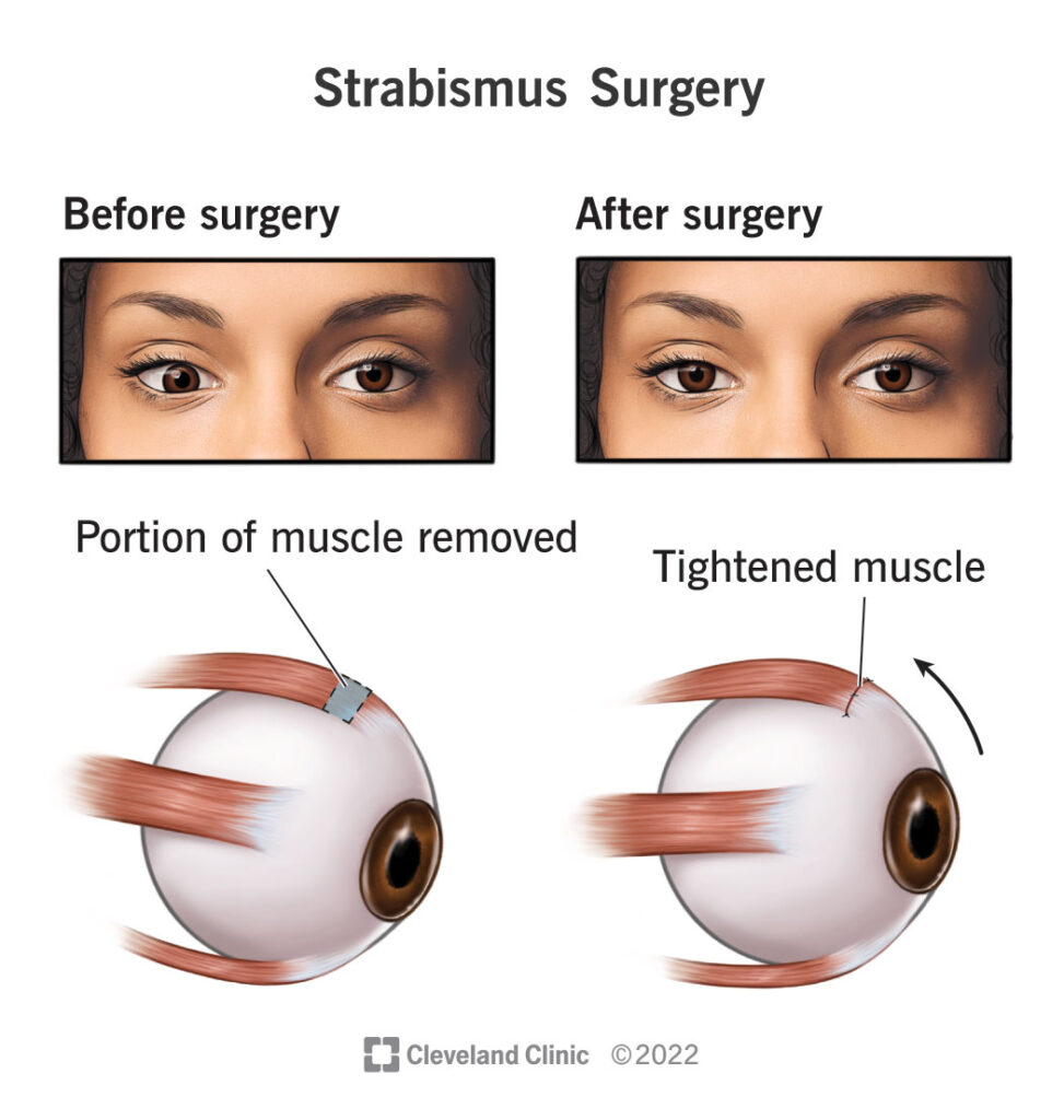
Strabismus Surgery
Involves tightening or weakening of extraocular muscles to correct misalignment.
Congenital Glaucoma Management
Goniotomy or trabeculotomy are preferred approaches in infants with buphthalmos and high IOP.
Complications and Risk Management in Ocular Surgery
Common Postoperative Risks
- Infection (endophthalmitis)
- Elevated IOP
- Corneal edema
- Retinal detachment
- Cystoid macular edema
Mitigation Strategies
- Sterile technique
- Preoperative antisepsis with povidone-iodine
- Judicious use of intraoperative antibiotics
- Close postoperative follow-up
Innovations in Eye Surgery: Robotics and Gene Therapy
Robotic-Assisted Microsurgery
Enables sub-micron precision for retinal or subretinal interventions; still in early adoption phase.
Gene Therapy and Cell Therapy
Used for inherited retinal diseases like Leber congenital amaurosis. Luxturna is the first FDA-approved ocular gene therapy.
Artificial Retina and Implants
The Argus II retinal prosthesis system offers partial vision restoration in patients with retinitis pigmentosa.
Surgical procedures on the eye proper encompass a wide spectrum of interventions—from anterior segment procedures like cataract extraction and corneal transplantation to complex posterior segment surgeries such as vitrectomy and retinal detachment repair. Success in these surgeries demands anatomical expertise, refined microsurgical skills, and access to modern technological advancements. Proper preoperative assessment, meticulous technique, and vigilant postoperative care are essential for achieving optimal outcomes in ocular surgery.

