Superficial basal cell carcinoma (sBCC) is the second most common subtype of basal cell carcinoma (BCC), the most prevalent form of skin cancer globally. Unlike nodular or infiltrative BCC, the superficial variant tends to present as flat, scaly patches, often resembling eczema or psoriasis, and primarily affects sun-exposed areas of the skin. Despite its slow progression and low metastatic potential, superficial BCC requires prompt diagnosis and effective treatment to prevent local tissue damage and recurrence.
Understanding the Pathophysiology of Superficial BCC
Superficial BCC originates from basal keratinocytes within the epidermis and exhibits proliferation along the dermoepidermal junction. Unlike other subtypes, sBCC typically remains within the superficial layers of the skin, characterized histologically by:
- Multifocal tumor nests confined to the epidermis and upper dermis
- Peripheral palisading of basal cells
- Retraction artifact between tumor islands and surrounding stroma
Ultraviolet (UV) radiation, particularly UVB exposure, induces DNA mutations, especially in the PTCH1 gene of the Hedgehog signaling pathway, initiating carcinogenesis.
Risk Factors Associated with Superficial BCC
Several factors contribute to the development of superficial basal cell carcinoma:
- Chronic sun exposure and use of tanning beds
- Fair skin phenotype (Fitzpatrick skin types I and II)
- Immunosuppression (organ transplant recipients, HIV-positive individuals)
- Previous history of nonmelanoma skin cancer
- Arsenic exposure or genetic conditions (e.g., basal cell nevus syndrome)
sBCC commonly appears on the trunk, upper limbs, or shoulders—areas that may not receive daily sun but are prone to intermittent, intense exposure.
Clinical Presentation and Symptoms
Superficial BCC typically presents as:
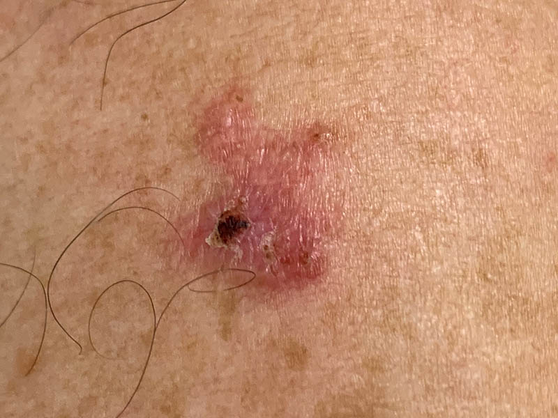
- Well-demarcated, erythematous patches or plaques
- Slightly elevated borders with central clearing
- Scaly, thin surface that may crust or bleed
- Asymptomatic or mildly pruritic
Unlike nodular BCC, which forms pearly papules, superficial BCC often mimics inflammatory dermatoses, delaying diagnosis if not evaluated clinically and histologically.
Diagnostic Methods for Superficial Basal Cell Carcinoma
Clinical Evaluation
- Dermoscopic examination reveals fine arborizing vessels, shiny white structures, and spoke-wheel areas.
- History of lesion persistence, growth, or non-response to topical corticosteroids should raise suspicion.
Skin Biopsy
- Punch biopsy is the preferred diagnostic tool to confirm histological subtype and exclude aggressive variants.
- Shave biopsy may be used for thin lesions with clear margins.
Histopathological Features
- Tumor nests in multifocal superficial arrangements
- Extension from the epidermis into the papillary dermis
- Peripheral palisading and mucinous stroma presence
Treatment Options for Superficial BCC
Management strategies vary depending on lesion size, location, cosmetic concern, and patient comorbidities.
1. Topical Therapies
Ideal for low-risk, superficial lesions:
- Imiquimod 5% cream: Immunomodulator applied 5 times per week for 6–12 weeks.
- 5-Fluorouracil (5-FU): Antimetabolite that disrupts DNA synthesis; applied twice daily for 3–6 weeks.
2. Surgical Interventions
Curettage and Electrodessication
- High success rate for superficial lesions
- Involves scraping the tumor and cauterizing the base
- Commonly used on the trunk and extremities
Excisional Surgery
- Preferred for larger or recurrent lesions
- 4–5 mm margin recommended
- Provides complete histological margin assessment
3. Photodynamic Therapy (PDT)
- Combines photosensitizing agents (aminolevulinic acid) with light activation
- Effective in cosmetically sensitive areas such as the face
- Multiple sessions may be required
4. Cryotherapy
- Involves rapid freezing with liquid nitrogen
- May be used for small, well-demarcated lesions
- Risk of hypopigmentation or scarring
Prognosis and Recurrence Rates
Superficial BCC is rarely life-threatening but may recur if inadequately treated. Prognostic factors include:
- Incomplete excision
- Immunosuppressed status
- Multifocal or extensive lesions
- Location on head and neck
Recurrence rates vary by treatment modality:
| Treatment Modality | Recurrence Rate (5 Years) |
|---|---|
| Surgical excision | 5–10% |
| Curettage & electrodessication | 7–15% |
| Imiquimod | 10–20% |
| Photodynamic therapy | 15–25% |
Prevention Strategies
Long-term management includes patient education and preventive care:
- Sun protection: Daily use of broad-spectrum SPF ≥30 sunscreen
- Regular skin exams: Self-exams and annual dermatologist visits
- Avoidance of tanning beds
- Vitamin D optimization through diet or supplements
High-risk individuals may benefit from chemopreventive agents such as nicotinamide.
Differential Diagnosis of Superficial BCC
Due to its subtle presentation, sBCC is often misdiagnosed. Conditions to consider include:
- Psoriasis
- Nummular eczema
- Lichen planus
- Actinic keratosis
- Bowen’s disease (squamous cell carcinoma in situ)
Dermoscopy and biopsy remain crucial in establishing a definitive diagnosis.
Follow-Up and Surveillance
Patients treated for superficial BCC require ongoing follow-up to detect recurrence or new lesions.
- Every 6–12 months for the first 2 years
- Annual skin exams thereafter
- Dermoscopic imaging for high-risk patients or multiple lesions
Early detection of recurrences improves outcomes and minimizes the need for aggressive treatments.
Superficial basal cell carcinoma is a prevalent but treatable form of nonmelanoma skin cancer. With accurate diagnosis and tailored therapy, outcomes are favorable. Proactive skin care, sun protection, and regular dermatological evaluations are essential in preventing recurrence and preserving skin health.