Streptococcal joint infection, a form of septic arthritis, is an acute and potentially destructive condition of the joints caused by Streptococcus species, most commonly Streptococcus pyogenes (Group A Streptococcus) and Streptococcus agalactiae (Group B Streptococcus). This infection invades the synovial membrane, leading to rapid joint degradation, intense inflammation, and systemic symptoms if not promptly treated.
Etiology: How Streptococcus Causes Joint Infections
Streptococcal joint infection typically occurs through hematogenous spread, direct inoculation from trauma or surgery, or contiguous spread from adjacent infected tissues. The bacteria enter the joint space and incite an inflammatory cascade involving neutrophil infiltration, synovial hyperplasia, and cartilage destruction.
Common Causative Agents
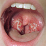
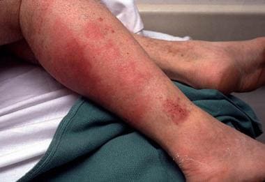
- Group A Streptococcus (GAS) – associated with pharyngitis and skin infections, but capable of invading joints
- Group B Streptococcus (GBS) – often involved in neonatal and elderly infections
- Streptococcus pneumoniae – linked with systemic bacteremia and immunocompromised patients
- Viridans streptococci – rare, but may cause infections in individuals with underlying joint disease or immunosuppression
Risk Factors for Streptococcal Joint Infection
Several underlying conditions and external factors increase susceptibility to joint infections:
- Pre-existing joint disease (e.g., rheumatoid arthritis, osteoarthritis)
- Recent joint surgery or injection
- Immunosuppression (HIV, chemotherapy, corticosteroids)
- Diabetes mellitus
- Hemodialysis or chronic renal failure
- Neonatal and geriatric age groups
- Skin infections or trauma near joints
Clinical Manifestations of Streptococcal Joint Infection
Streptococcal septic arthritis presents as a rapidly evolving monoarthritis, often involving large joints such as the knee, hip, or shoulder. Symptoms can include:
- Sudden onset of joint pain and swelling
- Restricted range of motion
- High-grade fever and chills
- Redness and warmth over the affected joint
- Systemic malaise and fatigue
In children and neonates, signs may be subtle, presenting with irritability, refusal to bear weight, or decreased limb movement.
Diagnostic Approach to Streptococcal Joint Infections
Clinical Examination
- Inspection and palpation for swelling, erythema, tenderness
- Joint immobility and passive motion pain
Laboratory Investigations
- Blood tests: Elevated white blood cells, ESR, and C-reactive protein (CRP)
- Blood cultures: May identify streptococcal bacteremia
- Synovial fluid analysis:
- Appearance: Turbid or purulent
- WBC count: Typically >50,000 cells/µL with neutrophilic dominance
- Gram stain and culture: Essential for definitive organism identification
- PCR assays: Useful for rapid pathogen detection
Imaging Studies
- X-rays: May show joint space narrowing or effusion in later stages
- Ultrasound: Useful for guiding joint aspiration
- MRI: Detects early bone and cartilage involvement, abscess formation
Treatment Protocols for Streptococcal Joint Infection
Empirical Antibiotic Therapy
Immediate initiation of intravenous antibiotics is critical before culture results are available. Empirical options include:
- Ceftriaxone or Cefotaxime – broad coverage including streptococci
- Vancomycin – in case of penicillin allergy or MRSA risk
Once streptococcus is confirmed and sensitivities are known:
- Penicillin G or Ceftriaxone remains the first-line choice
- Duration: Typically 2–4 weeks, beginning with IV administration, transitioning to oral antibiotics based on clinical response
Joint Drainage and Debridement
- Arthrocentesis: Daily or repeated aspiration of purulent fluid
- Arthroscopic lavage: Preferred for deeper joints like the hip
- Open surgical drainage: Reserved for unresponsive or complex cases
Supportive Measures
- Immobilization during the acute phase
- Physical therapy to restore mobility and prevent stiffness
- Analgesics and anti-inflammatory agents to manage pain
Prognosis and Potential Complications
When treated promptly, streptococcal joint infections carry a good prognosis. However, delayed intervention can lead to serious outcomes:
- Joint destruction and deformity
- Chronic osteomyelitis
- Functional disability
- Sepsis and multi-organ failure
- Growth plate damage in children
Prevention and Risk Mitigation
Preventive Measures
- Proper skin and wound care to avoid entry points for streptococcus
- Aseptic technique during joint injections and surgeries
- Vigilant management of skin infections near joints
- Early antibiotic treatment of streptococcal pharyngitis or cellulitis
Prophylactic Considerations
- Antibiotic prophylaxis in patients undergoing joint replacement with known history of invasive streptococcal disease
- Neonatal screening and intrapartum prophylaxis for Group B Streptococcus
Frequently Asked Questions:
How does streptococcus infect joints?
It typically reaches the joint through the bloodstream or from adjacent infected tissue, causing inflammation and damage.
Which joints are most commonly affected?
Large joints such as the knees, hips, and shoulders are most frequently involved.
Is streptococcal joint infection contagious?
While the joint infection itself is not contagious, the bacteria (e.g., Group A Strep) can spread via droplets or contact.
What is the duration of treatment for streptococcal joint infection?
Treatment generally lasts 2–4 weeks, with initial IV antibiotics followed by oral therapy.
Can streptococcal arthritis recur?
Recurrence is rare but possible, especially in immunocompromised individuals or those with underlying joint conditions.
Streptococcal joint infection is a serious medical condition requiring swift recognition and aggressive treatment. Early diagnosis via clinical assessment, synovial fluid analysis, and imaging is essential to prevent permanent joint damage. With appropriate antibiotic therapy and drainage, most patients recover fully, though delayed or inadequate treatment can result in irreversible disability. Maintaining vigilance in high-risk populations and adhering to aseptic practices during joint interventions remain key components in minimizing incidence and improving patient outcomes.