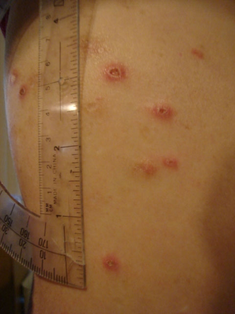
Skin ulcers are open sores or lesions resulting from poor blood circulation, prolonged pressure, or underlying medical conditions. These chronic wounds are a significant concern in both outpatient and inpatient care, particularly among the elderly, immobilized, and diabetic populations. Understanding their pathogenesis, risk factors, and management strategies is critical for timely intervention and optimized healing outcomes.

Understanding Skin Ulcers
A skin ulcer is defined as a breakdown in the skin and underlying tissue that fails to heal in a timely manner. It often develops due to insufficient blood flow, infection, or sustained mechanical stress. The affected area becomes necrotic, painful, and susceptible to secondary infections, further complicating recovery.
Key Causes of Skin Ulceration
1. Impaired Circulation
- Venous insufficiency: Leads to venous stasis ulcers, primarily in the lower legs.
- Arterial insufficiency: Causes ischemic ulcers due to blocked arteries and poor oxygen supply.
2. Diabetes Mellitus
- Peripheral neuropathy and poor wound healing contribute to diabetic foot ulcers.
3. Prolonged Pressure
- Extended immobility results in pressure ulcers (bedsores), especially in bedridden individuals.
4. Trauma and Injury
- Cuts, burns, or abrasions may progress into ulcers if infected or left untreated.
5. Infections
- Bacterial, fungal, or parasitic infections can compromise skin integrity and lead to ulcer formation.
Types of Skin Ulcers
Accurate classification informs prognosis and guides therapeutic choices. The major types include:
1. Venous Ulcers
- Location: Medial aspect of the lower leg
- Appearance: Shallow, irregular borders, weeping exudate, surrounded by hyperpigmentation
- Cause: Chronic venous insufficiency and valve incompetence
2. Arterial Ulcers
- Location: Toes, feet, lateral malleolus
- Appearance: Well-defined edges, dry or necrotic base, pale or cold skin
- Cause: Atherosclerosis and reduced arterial perfusion
3. Diabetic Foot Ulcers
- Location: Pressure points on feet, especially under the metatarsal heads
- Appearance: Painless, deep, with surrounding callus
- Cause: Neuropathy, trauma, poor glycemic control
4. Pressure Ulcers (Decubitus Ulcers)
- Location: Sacrum, heels, hips, shoulders
- Appearance: Staged from skin redness (Stage I) to exposed bone (Stage IV)
- Cause: Prolonged pressure, shear, and friction
5. Neurotrophic Ulcers
- Result from nerve damage and are similar to diabetic ulcers but can also be due to spinal cord injury or syphilis.
Clinical Symptoms of Skin Ulcers
- Persistent open sore
- Pain or tenderness (may be absent in neuropathic ulcers)
- Discolored or inflamed surrounding skin
- Foul-smelling discharge
- Swelling or warmth in the affected area
- Delayed healing over several weeks
Diagnostic Evaluation
1. Clinical Assessment
- Ulcer size, depth, location, exudate, and signs of infection
2. Laboratory Tests
- Blood glucose (for diabetic ulcers)
- Complete blood count and inflammatory markers
3. Imaging Studies
- Doppler ultrasound: Assess venous or arterial flow
- X-ray/MRI: Rule out osteomyelitis in deep or chronic ulcers
4. Wound Cultures
- To guide targeted antimicrobial therapy in infected ulcers
Treatment Strategies for Skin Ulcers
1. Wound Care Management
- Regular cleaning with sterile saline
- Debridement of necrotic tissue (surgical, enzymatic, autolytic)
- Application of dressings appropriate to wound type (hydrocolloids, alginates, foams)
2. Infection Control
- Topical antimicrobials (e.g., silver dressings)
- Systemic antibiotics for infected or cellulitic ulcers
3. Compression Therapy
- Especially effective for venous ulcers
- Use of gradient compression stockings or bandaging
4. Offloading and Pressure Relief
- For pressure and diabetic ulcers
- Use of special mattresses, cushions, and orthotic devices
5. Surgical Interventions
- Skin grafts or flaps for non-healing or extensive ulcers
- Revascularization for arterial insufficiency
6. Adjunct Therapies
- Negative Pressure Wound Therapy (NPWT)
- Hyperbaric Oxygen Therapy (HBOT)
- Growth factors and biologic dressings
Preventive Measures
1. Mobility and Repositioning
- Turn immobile patients every two hours
- Encourage ambulation when possible
2. Skincare and Hygiene
- Keep skin dry and moisturized
- Avoid harsh soaps and friction
3. Manage Underlying Conditions
- Control diabetes and hypertension
- Improve nutrition and hydration
4. Foot Care in Diabetics
- Routine foot inspections
- Proper footwear and podiatric consultations
Complications of Untreated Skin Ulcers
- Cellulitis
- Sepsis
- Osteomyelitis
- Chronic pain and disability
- Amputation in severe diabetic ulcers
- Malignancy (Marjolin’s ulcer)
Prognosis and Healing Timeline
- Healing depends on ulcer type, depth, comorbidities, and treatment adherence.
- Venous ulcers may take several months; diabetic and arterial ulcers often require long-term care.
- Early diagnosis and aggressive treatment dramatically improve outcomes.
Skin ulcers demand vigilant assessment and a multidisciplinary treatment approach. Addressing the root cause—whether vascular, metabolic, or mechanical—is essential to halt progression and promote healing. With structured wound care, prevention protocols, and patient education, we can significantly reduce ulcer-related morbidity and enhance quality of life.

