Sézary disease, also referred to as Sézary syndrome, is a rare and aggressive leukemic variant of cutaneous T-cell lymphoma (CTCL). This hematologic malignancy is marked by the presence of malignant T-lymphocytes—called Sézary cells—in the skin, lymph nodes, and peripheral blood. Distinguished from its less aggressive counterpart, mycosis fungoides, Sézary disease features systemic involvement and diffuse skin erythroderma, posing significant challenges in diagnosis and management.
Pathogenesis and Molecular Characteristics
Sézary disease originates from mature CD4+ memory T-cells with a skin-homing phenotype. These malignant lymphocytes display loss of normal surface antigens (such as CD7 and CD26) and harbor clonal T-cell receptor (TCR) gene rearrangements.
Key Pathophysiological Mechanisms
- Clonal expansion of atypical CD4+ T-cells
- Disruption of skin immunity leading to erythroderma
- Infiltration of lymph nodes and peripheral circulation
- Immune evasion via PD-1/PD-L1 and other checkpoint pathways
Epidemiology and Risk Factors
Sézary disease is an exceedingly rare condition with an incidence rate of approximately 0.3 cases per million annually. It typically affects adults over 60 years, with a slight male predominance. The etiology remains unclear, although potential contributing factors include:
- Genetic mutations (e.g., TP53, PLCG1, STAT3)
- Chronic immune stimulation
- Environmental exposures
- Prior cutaneous T-cell lymphoma (e.g., mycosis fungoides)
Clinical Presentation of Sézary Syndrome
Cardinal Symptoms
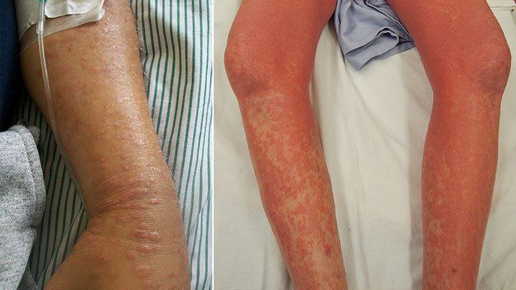
- Erythroderma: Diffuse reddening of ≥80% body surface area with scaling and pruritus
- Lymphadenopathy: Enlarged, sometimes painful, lymph nodes
- Sézary Cells: Malignant T-cells with cerebriform nuclei in blood
- Alopecia, nail dystrophy, and ectropion: Resulting from chronic skin inflammation
Systemic Features
- Fatigue
- Weight loss
- Recurrent infections due to immune dysregulation
Diagnostic Criteria for Sézary Disease
A definitive diagnosis requires the triad of:
- Erythroderma
- Clonal T-cell population in peripheral blood
- Presence of Sézary cells
Laboratory and Diagnostic Workup
- Complete blood count (CBC) with differential
- Flow cytometry: Shows loss of CD7, CD26 on CD4+ T-cells and increased CD4:CD8 ratio (>10)
- T-cell receptor gene rearrangement: PCR-based clonality testing
- Skin biopsy: Epidermotropism and atypical lymphocytes
- Lymph node biopsy: Confirms nodal involvement
- Imaging: CT or PET-CT for staging
Staging and Classification
The TNMB classification system is used to stage Sézary syndrome, evaluating:
- T (Skin involvement)
- N (Lymph node involvement)
- M (Visceral metastasis)
- B (Blood involvement)
Example:
- T4N3M0B2 = Generalized erythroderma, clinically abnormal lymphadenopathy, no visceral spread, and high Sézary cell count in blood.
Treatment Strategies for Sézary Disease
Treatment is individualized based on disease stage, patient health, and symptom severity. As Sézary syndrome is rarely curable, the goal is to manage symptoms and delay progression.
First-Line Therapies
- Photopheresis (ECP): Immunomodulatory therapy effective for blood and skin symptoms
- Interferon-alpha: Enhances immune response and reduces tumor burden
- Bexarotene (oral retinoid): Normalizes cell growth in malignant T-cells
Systemic Therapies
- Histone deacetylase (HDAC) inhibitors: Vorinostat, romidepsin for advanced disease
- Monoclonal antibodies:
- Mogamulizumab: Targets CCR4 on malignant T-cells
- Alemtuzumab: Anti-CD52 antibody for refractory cases
Advanced and Refractory Disease
- Allogeneic stem cell transplantation: Potentially curative in selected patients
- Combination chemotherapy: For rapidly progressive or life-threatening disease
Prognosis and Survival Rates
Sézary syndrome is associated with a poor prognosis due to its leukemic nature and systemic involvement.
- Median survival: 2–4 years from diagnosis
- Poor prognostic factors:
- Age >60
- High LDH levels
- Extensive blood involvement
- Poor response to initial therapy
Despite advances in targeted therapies, relapse is common, necessitating long-term surveillance and supportive care.
Supportive Care and Quality of Life
Patients often suffer from severe pruritus, skin breakdown, and psychosocial stress. Comprehensive care includes:
- Skin emollients and antihistamines
- Infection prevention and management
- Nutritional support
- Psychological counseling and social support services
Future Directions in Sézary Disease Research
Emerging therapies targeting immune checkpoints and molecular pathways offer hope for improved outcomes. Current investigational strategies include:
- PD-1/PD-L1 inhibitors
- CAR-T cell therapy targeting T-cell antigens
- JAK/STAT pathway inhibitors
- Tumor microenvironment modulation
Ongoing clinical trials are critical to defining the role of these agents in frontline and salvage settings.
FAQs
What is the difference between Sézary syndrome and mycosis fungoides?
Sézary syndrome is a leukemic form of CTCL involving the blood, whereas mycosis fungoides typically remains confined to the skin in early stages.
Are Sézary cells cancerous?
Yes, Sézary cells are malignant CD4+ T-lymphocytes characterized by cerebriform nuclei found in the blood and skin.
Is Sézary disease hereditary?
No clear hereditary link exists, although genetic mutations are implicated in its pathogenesis.
Can Sézary syndrome be cured?
It is rarely curable; however, long-term remission may be possible with stem cell transplantation or combination therapies.
How is Sézary disease different from eczema or psoriasis?
Unlike benign dermatoses, Sézary disease involves systemic symptoms, clonal T-cell proliferation, and malignant spread to blood and lymph nodes.