Severe ocular inflammation refers to intense inflammatory responses within the eye’s structures, including the uvea, sclera, retina, and optic nerve. Left untreated, this condition can lead to irreversible vision loss, ocular pain, and systemic complications. It commonly arises from autoimmune, infectious, or idiopathic etiologies, necessitating prompt and targeted intervention.
Common Causes of Severe Ocular Inflammation
Autoimmune and Systemic Inflammatory Diseases
- Behçet’s disease: Recurrent oral/genital ulcers, retinal vasculitis
- Sarcoidosis: Granulomatous uveitis with lung involvement
- Ankylosing spondylitis: HLA-B27 associated anterior uveitis
- Rheumatoid arthritis: Episcleritis or scleritis as a manifestation
Infectious Triggers
- Toxoplasmosis: Most common cause of posterior uveitis globally
- Tuberculosis: Granulomatous inflammation involving the choroid or retina
- Syphilis: “The great masquerader” affecting any ocular tissue
- Herpes viruses (HSV/VZV): Necrotizing retinitis, keratouveitis
Idiopathic Conditions
- Often classified after ruling out infectious and systemic causes
- Includes idiopathic panuveitis and white dot syndromes (e.g., birdshot chorioretinopathy)
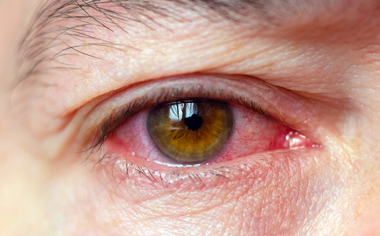
Clinical Presentation and Symptoms
- Eye pain: Sharp, deep orbital discomfort
- Redness: Particularly in anterior uveitis and scleritis
- Photophobia: Sensitivity to light due to ciliary spasm
- Blurred vision: Retinal or optic nerve involvement
- Floaters and visual field defects: Indicative of vitreous inflammation or retinal vasculitis
Severity and symptomatology vary based on the anatomical location and underlying etiology.
Diagnostic Approach for Severe Ocular Inflammation
Detailed Clinical Evaluation
- Slit-lamp examination: To assess anterior chamber cells, keratic precipitates, and iris synechiae
- Fundoscopy: Evaluation of vitreous haze, retinal lesions, vasculitis, or optic nerve swelling
Imaging Techniques
- Optical coherence tomography (OCT): Assesses retinal layers and macular edema
- Fluorescein angiography: Highlights vascular leakage or ischemia
- Ultrasound B-scan: Useful in dense vitritis or choroidal detachments
Laboratory and Systemic Workup
- Autoimmune markers: ANA, RF, HLA-B27, ACE, ANCA
- Infectious panels: VDRL/FTA-ABS (syphilis), Quantiferon (TB), toxoplasmosis titers
- Chest X-ray/CT: For sarcoidosis or tuberculosis suspicion
Management and Treatment Strategies
Corticosteroids: First-Line Anti-inflammatory Agents
- Topical steroids: Anterior segment inflammation (e.g., prednisolone acetate)
- Oral or IV steroids: Intermediate/posterior uveitis and severe cases
- Periocular injections: Sub-tenon’s triamcinolone for localized control
Immunosuppressive and Biologic Therapy
- Methotrexate, Azathioprine, Mycophenolate mofetil: For steroid-sparing long-term control
- Biologics:
- Adalimumab: FDA-approved for non-infectious uveitis
- Infliximab: Effective in Behçet’s-related uveitis and scleritis
Antimicrobial Therapy
- Tailored based on infectious etiology:
- Antitoxoplasma therapy: Pyrimethamine, sulfadiazine, and folinic acid
- Anti-TB regimens: Isoniazid, rifampin, ethambutol, pyrazinamide
- Antiviral agents: Acyclovir or valacyclovir for HSV/VZV
Surgical Interventions
- Vitrectomy: In cases of non-resolving vitritis, diagnostic sampling, or retinal detachment
- Cataract extraction: If vision-impairing cataract develops due to chronic inflammation or steroid use
- Glaucoma surgery: In steroid-induced or uveitic glaucoma cases
Complications of Untreated or Poorly Managed Inflammation
- Cystoid macular edema (CME)
- Secondary glaucoma
- Posterior synechiae and angle closure
- Retinal detachment
- Optic atrophy
- Permanent vision loss
Prompt treatment is essential to mitigate structural damage and preserve visual function.
Prognosis and Follow-up
Prognosis varies by cause, duration, and intensity of inflammation:
- Autoimmune causes: Often require lifelong immunomodulation
- Infectious etiologies: May have better outcomes if treated early
- Idiopathic cases: Require vigilant monitoring for recurrences
Follow-up Protocol
- Initial phase: Weekly to biweekly until inflammation subsides
- Maintenance phase: Monthly to quarterly, depending on disease activity and therapy
Preventive and Supportive Strategies
- Patient education: Emphasis on adherence and early symptom reporting
- Lifestyle modification: Sun protection, minimizing exposure to triggers
- Systemic disease coordination: Collaboration with rheumatologists or infectious disease specialists
Severe ocular inflammation constitutes a vision-threatening spectrum of disorders requiring urgent, multidisciplinary intervention. Comprehensive diagnostic evaluation, prompt initiation of therapy, and long-term follow-up are critical for preserving visual acuity and preventing irreversible ocular damage. As research advances, biologic therapies and personalized immunosuppression continue to improve outcomes for affected individuals.