Scleroderma, also known as systemic sclerosis, is a rare and chronic autoimmune connective tissue disease characterized by hardening and tightening of the skin and connective tissues. In some cases, it also affects internal organs such as the lungs, heart, kidneys, and gastrointestinal tract. The condition arises when the body’s immune system produces excess collagen, leading to tissue fibrosis, vascular abnormalities, and inflammation.
Types of Scleroderma and Their Clinical Features
Scleroderma is classified into two major types: localized scleroderma and systemic sclerosis, each with distinct manifestations and prognoses.
Localized Scleroderma
Primarily affects the skin and underlying tissues, without involving internal organs. It presents in two main forms:
- Morphea: Oval or linear patches of thickened skin that may fade over time.
- Linear Scleroderma: Bands of thickened skin typically seen in children, often on arms, legs, or forehead.
Systemic Sclerosis
Affects the skin, blood vessels, and internal organs, and is divided into:
- Limited Cutaneous Systemic Sclerosis (lcSSc): Skin thickening limited to the face and distal limbs; associated with CREST syndrome (Calcinosis, Raynaud’s, Esophageal dysmotility, Sclerodactyly, and Telangiectasia).
- Diffuse Cutaneous Systemic Sclerosis (dcSSc): Rapid progression of skin thickening involving trunk and proximal limbs with a higher risk of internal organ involvement.
Causes and Risk Factors of Scleroderma
The exact cause remains unknown, but multiple genetic, environmental, and immunologic factors contribute to disease onset.
Key Factors:
- Autoimmune dysfunction: The immune system mistakenly attacks healthy tissue, stimulating excessive collagen production.
- Genetics: Family history may play a role, though inheritance is uncommon.
- Environmental triggers: Exposure to silica dust, solvents, and certain viral infections may increase risk.
- Hormonal influence: Women are disproportionately affected, suggesting a hormonal component.
Symptoms and Signs of Scleroderma
The clinical presentation of scleroderma is highly variable and depends on the type and extent of organ involvement.
Common Dermatologic and Musculoskeletal Symptoms
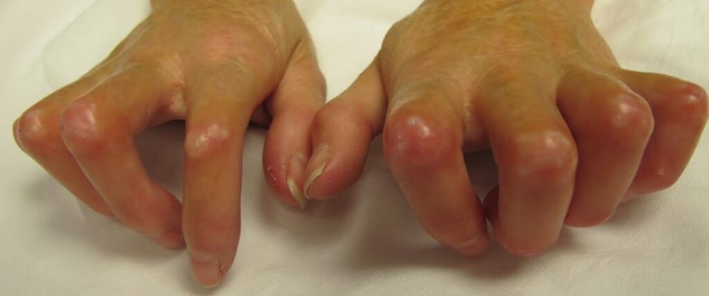
- Skin thickening and tightness, especially on the fingers and face
- Shiny, taut skin over joints
- Raynaud’s phenomenon: Whitening and blue discoloration of fingers or toes in response to cold
- Sclerodactyly: Thickened, claw-like fingers
- Calcinosis: Calcium deposits under the skin
- Joint pain and muscle weakness
Internal Organ Involvement
- Lungs: Interstitial lung disease, pulmonary arterial hypertension
- Heart: Arrhythmias, myocardial fibrosis
- Kidneys: Scleroderma renal crisis, potentially life-threatening
- GI tract: Esophageal reflux, dysphagia, malabsorption, constipation
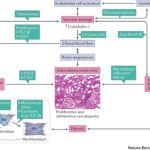
Pathophysiology of Scleroderma
The disease is marked by three central mechanisms:
This process disrupts the normal function of skin and organs by replacing healthy tissue with thickened, fibrotic tissue, leading to loss of elasticity, function, and vascular integrity.
Diagnostic Criteria and Tests
Scleroderma diagnosis is clinical but supported by laboratory tests and imaging to assess organ involvement.
Diagnostic Approach
- Medical history and physical examination
- Skin biopsy (in some cases) to confirm fibrosis
- Serological tests:
- ANA (antinuclear antibody): Present in most cases
- Anti-centromere antibodies: Common in lcSSc
- Anti-Scl-70 (anti-topoisomerase I): Associated with dcSSc
- Anti-RNA polymerase III: Linked with renal crisis risk
- Nailfold capillaroscopy: Reveals microvascular abnormalities
- Pulmonary function tests and HRCT: Assess interstitial lung disease
- Echocardiography: Evaluates pulmonary hypertension
- Renal function tests and urinalysis: Monitor for renal crisis
Differential Diagnosis: Distinguishing Scleroderma from Other Disorders
| Condition | Key Differences |
|---|---|
| Lupus erythematosus | Malar rash, renal disease, arthritis, photosensitivity |
| Dermatomyositis | Muscle weakness with skin rashes |
| Eosinophilic fasciitis | No Raynaud’s, eosinophilia prominent |
| Mixed connective tissue disease | Overlap of features, specific antibodies present |
Treatment Options for Scleroderma
There is no cure for scleroderma, but early, targeted therapy can mitigate symptoms, slow progression, and prevent complications.
Pharmacologic Treatments
- Immunosuppressants: Methotrexate, mycophenolate mofetil, cyclophosphamide (for skin and lung involvement)
- Corticosteroids: Use cautiously; can trigger renal crisis in high doses
- ACE inhibitors: First-line for scleroderma renal crisis
- Calcium channel blockers: For Raynaud’s phenomenon
- Proton pump inhibitors (PPIs): Manage esophageal reflux
- Endothelin receptor antagonists: Treat pulmonary arterial hypertension
Emerging Therapies
- Biologics: Tocilizumab, rituximab under investigation
- Autologous stem cell transplantation: Considered for severe, refractory cases
Lifestyle Modifications and Supportive Care
Managing scleroderma requires a multidisciplinary approach and lifestyle adjustments.
- Protect skin from trauma and cold to reduce Raynaud’s symptoms
- Moisturizers and gentle skincare routines
- Small, frequent meals to manage gastrointestinal symptoms
- Physical therapy to maintain joint flexibility and muscle strength
- Psychological support for emotional well-being
- Smoking cessation to improve circulation
Prognosis and Complications
Prognosis varies by subtype and extent of organ involvement.
Favorable Prognosis:
- Limited cutaneous form
- Absence of internal organ complications
- Early and consistent treatment
Poor Prognosis Indicators:
- Rapid skin thickening
- Pulmonary arterial hypertension
- Scleroderma renal crisis
- Cardiac involvement
Scleroderma is a complex, multisystem autoimmune disorder with significant clinical variability. Through early diagnosis, organ-specific treatment, and coordinated care, individuals can manage symptoms effectively and maintain quality of life. Ongoing research into biologic agents and disease-modifying therapies continues to offer new hope for improved outcomes.