Retinal vascular occlusion is a critical ophthalmologic condition resulting from blockage in the retinal arteries or veins. It is often referred to as an “eye stroke” due to its sudden onset and potential for rapid vision loss. As a leading cause of irreversible visual impairment, timely recognition and intervention are essential.
This condition encompasses two primary subtypes:
- Retinal Artery Occlusion (RAO)
- Retinal Vein Occlusion (RVO)
Both forms share overlapping risk factors but differ significantly in pathophysiology, presentation, and treatment strategies.
Classification of Retinal Vascular Occlusion
Retinal Artery Occlusion (RAO)
RAO typically results from embolic or thrombotic events leading to ischemia in the retinal tissues. It is classified into:
- Central Retinal Artery Occlusion (CRAO)
- Branch Retinal Artery Occlusion (BRAO)
Retinal Vein Occlusion (RVO)
RVO is due to venous thrombosis or compression and leads to retinal hemorrhage and edema. It is classified into:
- Central Retinal Vein Occlusion (CRVO)
- Branch Retinal Vein Occlusion (BRVO)
Etiology and Risk Factors
Several systemic and ocular risk factors contribute to retinal vascular occlusions:
- Hypertension
- Diabetes mellitus
- Hyperlipidemia
- Atherosclerosis
- Cardiac arrhythmias or valvular disease
- Hypercoagulable states
- Glaucoma (notably with CRVO)
Patients with these conditions are predisposed to thromboembolic phenomena, emphasizing the need for systemic evaluation.
Clinical Presentation
The hallmark of retinal vascular occlusion is painless, sudden vision loss. Specific features vary by type:
Central Retinal Artery Occlusion (CRAO)
- Sudden, profound vision loss in one eye
- A “cherry red spot” at the macula on fundoscopic exam
- Retinal whitening and arteriolar attenuation
Branch Retinal Artery Occlusion (BRAO)
- Sectoral vision loss (e.g., visual field defects)
- Similar fundoscopic findings in a localized area
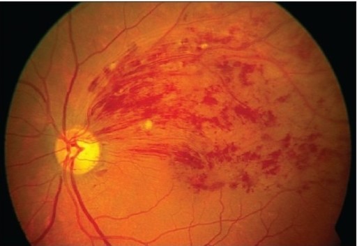
Central Retinal Vein Occlusion (CRVO)
- Blurred or distorted vision, typically unilateral
- Diffuse retinal hemorrhages (“blood and thunder” appearance)
- Optic disc edema and venous tortuosity
Branch Retinal Vein Occlusion (BRVO)
- Vision loss in a quadrant or sector
- Hemorrhages and edema confined to a specific region
Diagnostic Workup
Prompt diagnosis involves clinical evaluation supported by imaging.
Ophthalmologic Examination
- Visual acuity testing
- Amsler grid for central distortion
- Dilated fundus examination
Imaging Studies
- Optical Coherence Tomography (OCT): Detects macular edema and retinal layer integrity
- Fluorescein Angiography: Assesses perfusion, nonperfusion, and leakage
- OCT-Angiography: Noninvasive assessment of retinal vasculature
Systemic Evaluation
- Blood pressure and glucose monitoring
- Lipid panel and coagulation profile
- Cardiac echocardiography
- Carotid Doppler ultrasound
Management Strategies
Treatment is tailored to the type of occlusion and associated complications.
Retinal Artery Occlusion
RAO is a medical emergency. Interventions include:
- Ocular massage to dislodge emboli
- Anterior chamber paracentesis to reduce intraocular pressure
- Carbogen inhalation or hyperbaric oxygen therapy
- Thrombolytic therapy in select cases (intra-arterial tPA)
Outcomes depend heavily on the timing of presentation—vision recovery is rare if treatment is delayed beyond 4–6 hours.
Retinal Vein Occlusion
The management of RVO focuses on reducing macular edema and preventing neovascular complications:
- Intravitreal anti-VEGF injections (e.g., ranibizumab, aflibercept)
- Intravitreal corticosteroids (e.g., dexamethasone implants)
- Panretinal photocoagulation (PRP) for ischemic CRVO with neovascularization
- Management of systemic risk factors
Prognosis and Complications
Retinal Artery Occlusion
- Generally poor visual prognosis
- CRAO often leads to severe permanent vision loss
- BRAO may retain some visual field function
Retinal Vein Occlusion
- Prognosis varies with extent of ischemia
- CRVO carries a higher risk of neovascular glaucoma
- Macular edema is the leading cause of vision impairment
Regular follow-up is crucial to monitor for complications such as:
- Neovascularization of the retina or iris
- Macular ischemia
- Retinal detachment
- Secondary glaucoma
Preventive Measures
Preventing retinal vascular occlusion involves aggressive control of systemic conditions:
- Blood pressure control
- Glycemic management in diabetics
- Lipid-lowering therapy
- Antiplatelet agents for vascular risk
- Smoking cessation
- Regular ophthalmic checkups in high-risk populations
Collaboration with internists, cardiologists, and neurologists is vital to prevent recurrent events and systemic complications such as stroke.
Frequently Asked Questions:
What causes retinal vascular occlusion?
It is caused by blockage in the retinal arteries or veins due to emboli, thrombosis, or vascular compression.
Is vision loss from retinal vascular occlusion permanent?
It can be permanent, especially in artery occlusions. Prompt treatment improves chances of recovery in some cases.
How is it different from a stroke?
Although called an “eye stroke,” retinal vascular occlusion affects vision specifically but shares similar underlying risk factors with brain stroke.
What is the best treatment for vein occlusion in the eye?
Intravitreal anti-VEGF injections are the gold standard for managing macular edema in vein occlusion.
Can retinal vascular occlusion be prevented?
Yes, through control of blood pressure, cholesterol, diabetes, and avoiding smoking.