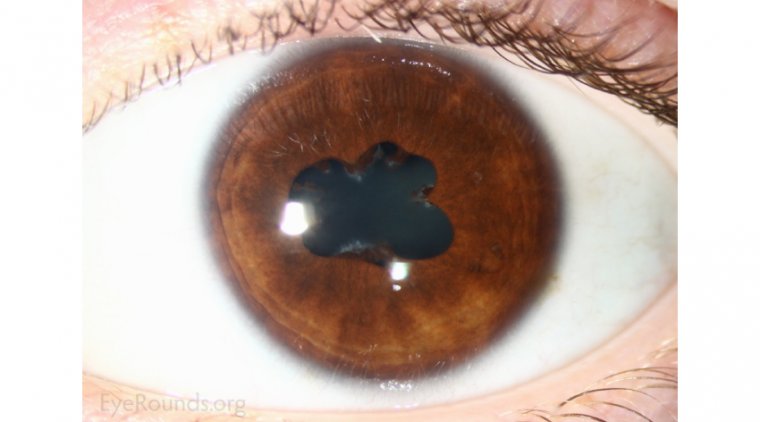
Posterior synechiae are adhesions between the posterior surface of the iris and the anterior lens capsule. These adhesions typically develop as a consequence of intraocular inflammation, most commonly in the context of anterior uveitis. If not promptly diagnosed and managed, posterior synechiae may lead to complications such as elevated intraocular pressure, angle-closure glaucoma, and permanent vision impairment.
Etiology: Causes and Risk Factors for Posterior Synechiae
The formation of posterior synechiae is most frequently associated with inflammatory ocular disorders. Inflammatory cells and fibrinous exudates promote adhesion between the iris and the lens.
Primary Causes:
- Anterior uveitis (iritis): The most common underlying cause
- Trauma: Direct or surgical trauma to the eye
- Infectious uveitis: Including herpes simplex virus, syphilis, tuberculosis
- Autoimmune conditions: Such as HLA-B27-associated uveitis, sarcoidosis, or juvenile idiopathic arthritis
Iatrogenic and Medication-Induced Causes:
- Prolonged use of miotics (e.g., pilocarpine)
- Post-surgical inflammation (e.g., cataract surgery without proper prophylaxis)
Pathophysiology of Iris-Lens Adhesions
During ocular inflammation, inflammatory mediators and fibrin are released into the aqueous humor, creating a sticky interface that binds the posterior iris to the lens. This adhesion can range from small focal points to extensive 360-degree involvement.
Consequences of Unresolved Synechiae:
- Pupillary block: Prevents normal aqueous flow, raising intraocular pressure
- Seclusio pupillae: Complete circular adhesion causing total blockage
- Secondary angle-closure glaucoma
- Permanent pupillary distortion or irregularity
Clinical Presentation and Symptoms
While some patients with posterior synechiae may remain asymptomatic, others exhibit noticeable ocular discomfort or visual changes.
Common Symptoms:
- Blurred vision
- Eye redness and pain
- Photophobia
- Irregularly shaped or fixed pupil
- Decreased visual acuity
- Halos around lights (if intraocular pressure rises)
Diagnosis and Examination Techniques
A thorough ophthalmic evaluation is essential for identifying posterior synechiae. Clinical signs may be subtle, especially in early stages.
Diagnostic Tools:
- Slit Lamp Biomicroscopy: Visualization of adhesions between the iris and lens
- Pupil Dilation Test: Poor or segmental dilation suggests synechial attachment
- Tonometry: Checks for elevated intraocular pressure
- Anterior Segment Optical Coherence Tomography (AS-OCT): Offers cross-sectional imaging of anterior chamber structures
Differentiating Posterior from Anterior Synechiae
| Feature | Posterior Synechiae | Anterior Synechiae |
|---|---|---|
| Location | Between iris and lens | Between iris and cornea |
| Associated Condition | Anterior uveitis | Chronic angle-closure glaucoma |
| Risk of Pupillary Block | High | Low |
| Management Strategy | Cycloplegics, steroids | Peripheral iridectomy |
Management of Posterior Synechiae
Treatment focuses on reducing inflammation, preventing further adhesions, and restoring normal pupil function. Early intervention improves the prognosis significantly.
First-Line Therapeutics:
- Topical corticosteroids: Prednisolone acetate 1% to control inflammation
- Cycloplegic agents: Atropine or homatropine to dilate the pupil and prevent further adhesion
- Mydriatics: Phenylephrine to break recent adhesions
Advanced Interventions:
- Synechiolysis: Manual or laser-assisted separation of adhesions
- Surgical intervention: Indicated in refractory cases or when accompanied by pupillary block and glaucoma
- Antiviral or antimicrobial therapy: If infectious uveitis is the cause
Preventive Measures in At-Risk Patients
Prevention is critical in individuals prone to recurrent uveitis or undergoing intraocular surgery.
Recommendations:
- Prophylactic use of cycloplegics and steroids during ocular procedures
- Close monitoring of patients with autoimmune uveitis
- Prompt treatment of any anterior segment inflammation
- Avoidance of long-term miotic therapy in susceptible patients
Long-Term Prognosis and Monitoring
The visual prognosis in patients with posterior synechiae depends on the extent of adhesion and the timeliness of intervention. Permanent pupillary distortion can occur, but severe visual loss is preventable with appropriate care.
Follow-up Considerations:
- Regular intraocular pressure monitoring
- Periodic dilated fundus examinations
- Monitoring for recurrence in chronic uveitis
- Adjusting therapy based on inflammatory activity and response
Posterior synechiae represent a critical ophthalmologic condition resulting from intraocular inflammation. Timely recognition and aggressive management are key to preventing irreversible complications like pupillary block and glaucoma. A structured approach involving anti-inflammatory therapy, pupil dilation, and when necessary, surgical intervention, ensures the best outcomes for affected individuals.