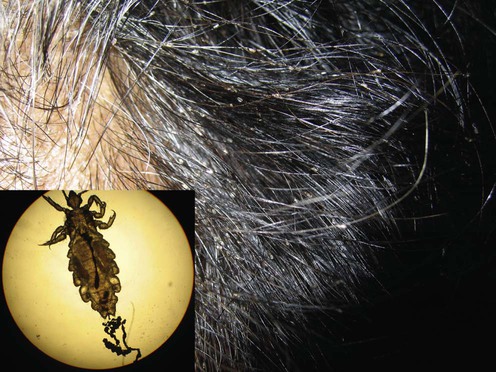
Pediculosis capitis, commonly known as head lice infestation, is a widespread parasitic condition caused by Pediculus humanus capitis. This obligate ectoparasite affects the scalp and hair shafts, predominantly in children between the ages of 3 and 12. Despite being non-life-threatening, it can cause significant discomfort, social stigma, and secondary infections if left untreated.
Epidemiology and Transmission of Pediculosis Capitis
Pediculosis capitis is highly contagious and primarily spreads through direct head-to-head contact. Though less common, transmission can also occur via fomites such as combs, hats, and bedding.
- Global prevalence: Varies from 1% to 40% in school-aged children
- Higher risk groups: Children in crowded conditions, especially in schools or daycare centers
- No association with hygiene: Lice infestation affects individuals irrespective of personal cleanliness
Life Cycle of Pediculus Humanus Capitis
Understanding the louse’s life cycle is critical for effective management. The cycle consists of three stages:
- Egg (Nit): Oval, yellowish-white, cemented to the hair shaft close to the scalp. Hatches in 7–10 days.
- Nymph: Immature louse that matures within 9–12 days after hatching.
- Adult: Lives up to 30 days on the scalp and lays up to 10 eggs per day.
Clinical Features of Head Lice Infestation
The primary symptoms result from the louse’s feeding behavior, as it pierces the scalp to consume human blood.
- Intense scalp itching (pruritus)
- Excoriations and secondary bacterial infections from scratching
- Tickling or crawling sensation in the hair
- Presence of nits, especially behind the ears and at the nape of the neck
- Regional lymphadenopathy in severe cases
- Insomnia and irritability in children
Diagnostic Approach to Pediculosis Capitis
A thorough visual examination remains the cornerstone of diagnosis. Tools and techniques include:
Visual Inspection:
- Use of fine-toothed lice comb on wet or conditioned hair increases detection sensitivity.
- Eggs located within 6 mm of the scalp indicate active infestation.
- Viable nits are usually tan to brown, while nonviable ones appear white.
Microscopy:
- Confirms diagnosis by identifying live lice or viable eggs
- Nits can be differentiated from dandruff or hair debris by their firm adherence to the hair shaft
Dermoscopy:
- Enhances visualization of eggs and lice in situ
- Useful in differentiating between viable and empty nits
Treatment of Pediculosis Capitis
An effective treatment plan targets live lice and viable eggs, while also preventing reinfestation.
First-Line Pharmacological Agents
| Treatment | Mechanism | Application | Comments |
|---|---|---|---|
| Permethrin 1% | Neurotoxic to lice | Applied to damp hair and left for 10 min | Most commonly used |
| Malathion 0.5% | Cholinesterase inhibitor | Applied to dry hair for 8–12 hrs | Flammable, use with caution |
| Dimethicone | Physical suffocation | Coats and dehydrates lice | Non-toxic, safe for children |
Second-Line and Alternative Agents
- Ivermectin (topical or oral) – effective against resistant lice
- Spinosad – kills lice and eggs with one application
- Benzyl alcohol 5% – asphyxiates lice but not ovicidal, requires repeat use
Non-Pharmacological and Mechanical Approaches
- Wet combing with conditioner every 3–4 days for 2 weeks
- Manual nit removal helps prevent recurrence
- Heat-based devices (e.g., LouseBuster) kill lice through dehydration
Ineffective or Discouraged Treatments
- Home remedies (e.g., mayonnaise, vinegar) lack scientific backing
- Insecticide misuse can lead to toxicity without improved efficacy
Prevention and Control Strategies
Personal and Environmental Measures
- Avoid head-to-head contact, especially among children
- Do not share personal items such as brushes, hats, and headphones
- Wash bedding and clothing in hot water (≥55°C)
- Seal non-washable items in plastic bags for 2 weeks
School and Public Health Policy
- Children should not be excluded from school due to lice
- The “no-nit policy” is discouraged by major health bodies (AAP, CDC)
- Education of families, teachers, and caregivers plays a key role
Management of Recurrent or Resistant Pediculosis
Re-infestation may occur due to:
- Inadequate treatment application
- Failure to repeat application for ovicidal agents
- Close contacts remaining untreated
- Insecticide resistance
Recommended Action:
- Reassess treatment technique and adherence
- Switch to a different agent with a different mechanism of action
- Treat close contacts and environment simultaneously
Complications of Untreated Pediculosis Capitis
Though rare, complications may include:
- Secondary bacterial infection: impetigo, cellulitis
- Lymphadenitis due to chronic inflammation
- Psychosocial stress and sleep disturbances in children
- Iron deficiency anemia in cases of severe chronic infestation
Global and Societal Impact
Despite the benign nature of the condition, pediculosis capitis has a significant societal burden, particularly in school systems where outbreaks can disrupt learning and prompt unnecessary absenteeism.
- Economic cost includes treatment, missed school/workdays, and doctor visits
- Stigma may cause embarrassment and emotional distress, especially in children
- Educational campaigns should focus on early detection and rational management rather than punitive isolation
Pediculosis capitis remains a pervasive pediatric concern globally. With appropriate diagnosis, effective treatment, and strategic prevention, this infestation can be controlled with minimal disruption to a child’s health or education. Modern pharmacologic and non-pharmacologic interventions offer safe and reliable results when applied correctly, especially when integrated with community-based education and hygiene practices.