Paucibacillary leprosy (PB leprosy) is a clinically and microbiologically distinct form of Hansen’s disease characterized by a limited number of skin lesions and low bacterial load. Recognizing this presentation promptly and managing it effectively is critical to halting transmission, preventing disability, and ensuring early cure.
Overview of Leprosy and Its Classification
Leprosy, caused by Mycobacterium leprae, manifests along a spectrum based on the host’s immune response. The World Health Organization (WHO) classifies leprosy into two major clinical forms for treatment purposes:
- Paucibacillary (PB) Leprosy: Fewer than five skin lesions, no detectable bacilli on skin smears
- Multibacillary (MB) Leprosy: Five or more lesions, or any number with positive skin smears
This operational classification simplifies diagnosis and ensures appropriate multidrug therapy (MDT) implementation.
Defining Features of Paucibacillary Leprosy
Clinical Presentation
PB leprosy is generally characterized by:
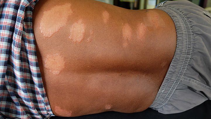
- 1 to 5 hypopigmented or erythematous skin lesions
- Well-defined or vague borders
- Loss of sensation over the affected area
- Thickened peripheral nerves, though less commonly than in MB cases
- No bacilli detected on slit-skin smears (negative smear)
These lesions may be dry, hairless, and devoid of sweating. Their asymmetry and non-pruritic nature often distinguish them from other dermatological conditions.
Pathogenesis and Immunological Response
In PB leprosy, the host mounts a strong cell-mediated immune response against M. leprae, restricting bacillary proliferation. This explains the absence of widespread lesions and negative bacteriological indices.
PB leprosy typically corresponds to tuberculoid or borderline tuberculoid forms on the Ridley-Jopling scale.
Diagnostic Criteria for Paucibacillary Leprosy
Diagnosis is primarily clinical, supported by:
- Skin lesion count (≤5)
- Loss of sensation over lesions
- Absence of acid-fast bacilli in slit-skin smears
Skin biopsy may aid in confirmation, revealing granulomatous inflammation with epithelioid cells and Langhans giant cells.
Essential Diagnostic Tools
| Diagnostic Tool | Utility |
|---|---|
| Clinical skin examination | Primary method |
| Slit-skin smear | Rule out multibacillary leprosy |
| Skin biopsy | Histological confirmation |
| Nerve function assessment | Detect early neuropathy |
Treatment Protocol for Paucibacillary Leprosy
WHO-Recommended Multidrug Therapy (MDT)
Treatment is both effective and standardized globally under the WHO program:
- Rifampicin 600 mg once monthly (supervised)
- Dapsone 100 mg daily (self-administered)
- Duration: 6 months
Key Considerations
- Adherence is essential to avoid relapse or resistance.
- Treatment is free in many endemic countries, encouraging early intervention.
- Patients should be monitored for drug reactions and nerve function impairment.
Monitoring and Managing Complications
Though PB leprosy has a limited bacterial load, nerve involvement may lead to:
- Numbness
- Muscle weakness
- Ulcers or deformities if untreated
Early recognition and corticosteroid therapy (e.g., prednisolone) are used to manage Type 1 lepra reactions involving nerve inflammation.
Public Health Strategies and Prevention
Contact Tracing and Chemoprophylaxis
- Close contacts of patients are at risk; periodic screening is advised.
- Single-dose rifampicin (SDR) is recommended for eligible contacts to reduce incidence.
Stigma Reduction and Awareness
Public education campaigns remain vital in reducing social stigma and promoting early self-reporting.
Differential Diagnosis
Paucibacillary leprosy may mimic several dermatological conditions, such as:
- Tinea versicolor
- Vitiligo
- Psoriasis
- Lichen planus
- Post-inflammatory hypopigmentation
Proper sensory testing and lesion evaluation are crucial for distinguishing PB leprosy from these conditions.
Prognosis and Follow-Up
With timely and complete MDT, the prognosis for PB leprosy is excellent. Patients are considered non-infectious after the first dose of treatment. Regular follow-up is necessary to:
- Monitor nerve function
- Identify relapse or late complications
- Reinforce adherence to treatment
Paucibacillary leprosy represents a curable and non-contagious form of Hansen’s disease with limited lesions and negative smear findings. Prompt diagnosis, standard WHO multidrug therapy, and vigilant follow-up are key to preventing disability and eliminating disease burden at the community level.