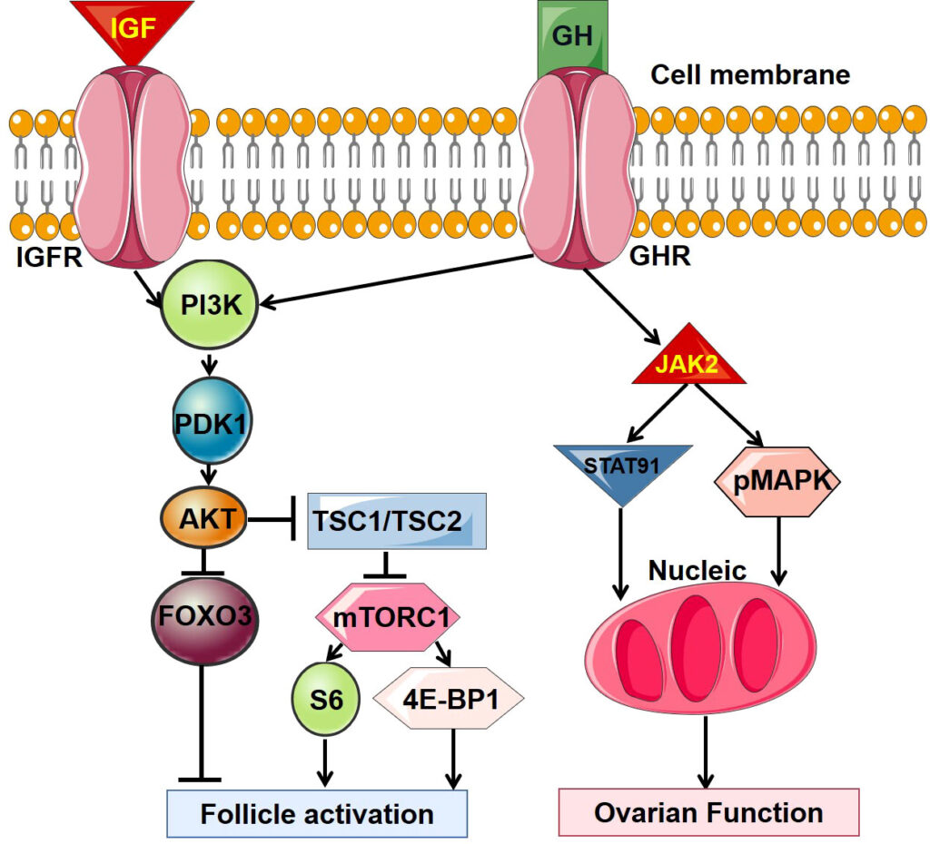
Ovarian function studies represent a crucial component of reproductive endocrinology, offering a structured evaluation of ovarian health, hormonal balance, and reproductive potential. These diagnostic tools are indispensable for assessing fertility, predicting menopausal onset, and guiding interventions in disorders such as polycystic ovary syndrome (PCOS), premature ovarian insufficiency (POI), and other ovulatory dysfunctions. By analyzing endocrine markers and imaging findings, we can gain an accurate understanding of ovarian reserve, cyclic function, and response to fertility treatments.

Hormonal Basis of Ovarian Function
The ovary is regulated by the hypothalamic-pituitary-ovarian (HPO) axis through a tightly controlled endocrine feedback loop:
Key Hormones in Ovarian Function:
- Follicle-Stimulating Hormone (FSH): Stimulates follicular growth.
- Luteinizing Hormone (LH): Triggers ovulation and corpus luteum formation.
- Estradiol (E2): Produced by developing follicles, regulates endometrial proliferation.
- Progesterone: Secreted by corpus luteum, essential for luteal phase maintenance.
- Anti-Müllerian Hormone (AMH): Secreted by preantral and small antral follicles, reflects ovarian reserve.
Ovarian Reserve Testing: Core of Ovarian Function Studies
Ovarian reserve indicates the quantity and quality of a woman’s remaining oocytes. The following tests are central to this assessment:
1. Anti-Müllerian Hormone (AMH)
- Significance: Independent of the menstrual cycle, AMH is the most stable and reliable marker.
- Interpretation:
- 3.5 ng/mL: High reserve (may indicate PCOS)
- 1.0–3.5 ng/mL: Normal reserve
- <1.0 ng/mL: Diminished reserve
2. Antral Follicle Count (AFC)
- Method: Transvaginal ultrasound to count follicles 2–10 mm in diameter on cycle days 2–5.
- Normal Range: 10–20 total follicles; <5 suggests diminished reserve.
3. Baseline FSH and Estradiol
- Timing: Measured on day 2–4 of the cycle.
- High FSH (>10 IU/L): Indicative of poor ovarian reserve.
- High Estradiol (>80 pg/mL): May falsely suppress FSH, masking true reserve status.
Additional Hormonal Assessments in Ovarian Function Studies
Luteinizing Hormone (LH)
- Elevated LH/FSH ratio (>2:1): Suggestive of PCOS.
Progesterone
- Mid-luteal Level (>3 ng/mL): Confirms ovulation.
Inhibin B
- Function: Reflects granulosa cell function.
- Use: Less commonly used but can support FSH interpretation.
Imaging Modalities in Ovarian Evaluation
Transvaginal Ultrasound
Provides direct visualization of ovarian volume, antral follicles, and stromal characteristics.
- Ovarian Volume:
- Normal: 3–10 cm³
- Increased in PCOS
- Decreased in ovarian insufficiency
Doppler Studies
Evaluate stromal blood flow to predict response in assisted reproductive technology (ART) cycles.
Functional Tests of Ovarian Responsiveness
Clomiphene Citrate Challenge Test (CCCT)
- Protocol: FSH measured on day 3, followed by clomiphene (100 mg/day on days 5–9), and FSH measured again on day 10.
- Elevated FSH on either day: Suggests reduced ovarian reserve.
Exogenous FSH Ovarian Reserve Test (EFORT)
- FSH administered on day 3; estradiol response assessed on day 4.
- Low E2 response: Indicative of poor ovarian responsiveness.
Applications of Ovarian Function Studies in Clinical Practice
Infertility Workup
- Purpose: To determine appropriate fertility interventions, including ovulation induction or IVF.
- Significance: Tailoring protocols based on ovarian reserve improves outcomes and reduces risks.
Assessment Prior to Cancer Treatment
- Fertility Preservation: AMH and AFC guide oocyte or embryo cryopreservation decisions.
Menopausal Transition
- Prediction of Menopause: Declining AMH levels signal approaching menopause.
- Hormone Therapy Decisions: Hormonal profiling assists in determining suitability for HRT.
Diagnosis of PCOS and POI
- PCOS Indicators: High AMH, high LH/FSH ratio, polycystic ovarian morphology.
- POI Indicators: Low AMH, high FSH, amenorrhea before age 40.
Interpretation and Limitations
Factors Affecting Accuracy
- Age: Natural decline in ovarian function starts in early 30s.
- Cycle Variability: Hormonal levels can fluctuate between cycles.
- Medications: Hormonal contraceptives may suppress AMH and AFC.
Complementary Use
No single test suffices. Combining AMH, AFC, and hormonal profiling offers the most robust diagnostic power.
Frequently Asked Questions:
What is the best time to test ovarian function?
Day 2–5 of the menstrual cycle is ideal for hormonal and ultrasound-based assessments.
Can ovarian function be restored?
While aging cannot be reversed, some interventions (e.g., DHEA supplementation or lifestyle changes) may improve function in certain conditions.
Does a low AMH mean infertility?
Not necessarily. AMH reflects quantity, not quality. Many women with low AMH still conceive naturally.
Are ovarian function studies painful?
Most are minimally invasive. Blood draws and ultrasounds involve little to no discomfort.
How often should ovarian function be evaluated?
In fertility planning or treatment, assessments are typically annual or cycle-based depending on the condition.
Ovarian function studies provide a foundational framework for evaluating reproductive potential and guiding fertility treatments. Through a detailed assessment of hormonal markers, ultrasound imaging, and functional tests, clinicians can accurately diagnose and manage diverse ovarian disorders. As precision medicine evolves, individualized interpretation of these studies becomes critical in optimizing reproductive outcomes and enhancing patient care.