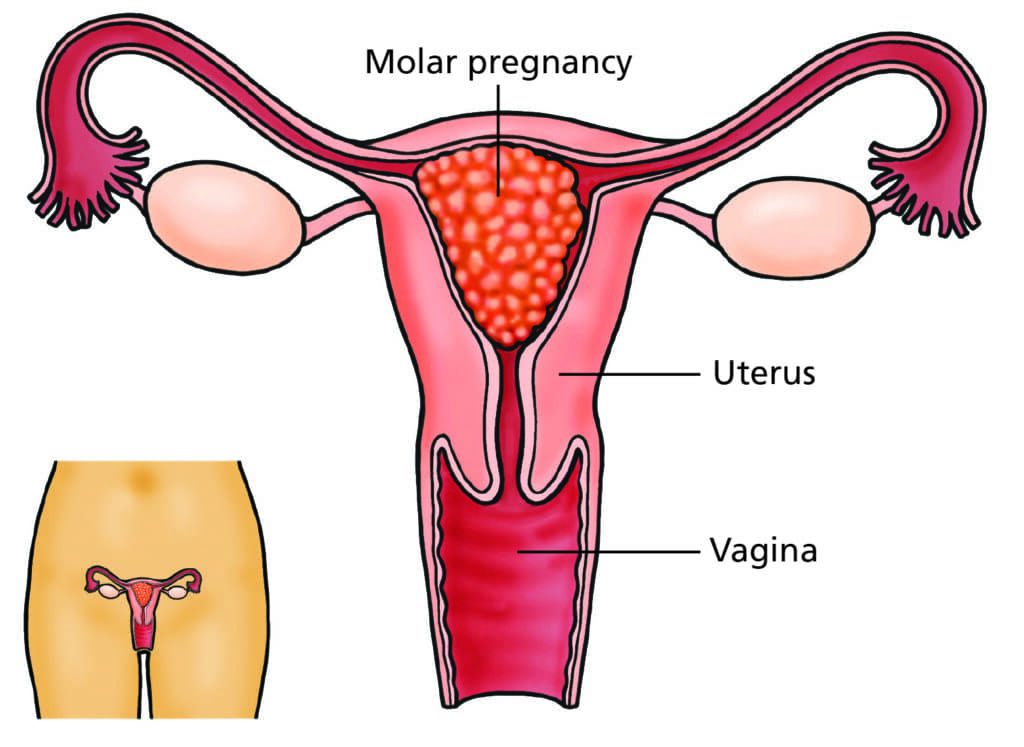
A benign hydatidiform mole, commonly referred to as a molar pregnancy, is a rare yet significant condition that can occur during pregnancy. This article delves into the details of benign hydatidiform mole, exploring its causes, symptoms, diagnostic approaches, and available treatment options. By gaining a thorough understanding of this condition, individuals can make informed decisions regarding their health and well-being.

What Is a Benign Hydatidiform Mole?
A benign hydatidiform mole is a type of gestational trophoblastic disease (GTD), a group of rare conditions that involve abnormal growth of the cells that would normally form the placenta during pregnancy. Unlike a typical pregnancy, where the fertilized egg develops into an embryo, a molar pregnancy results in the development of abnormal tissue within the uterus instead of a viable fetus.
Types of Hydatidiform Moles
There are two primary types of hydatidiform moles:
- Complete Hydatidiform Mole (CHM): In this form, no normal fetal tissue develops. Instead, the placenta forms a mass of cyst-like structures that can grow and spread.
- Partial Hydatidiform Mole (PHM): In a partial mole, some normal fetal tissue may develop alongside the abnormal placental tissue. However, the pregnancy is not viable.
While a complete hydatidiform mole is typically considered benign, it has the potential to progress into more severe complications if left untreated.
Causes of Benign Hydatidiform Mole
The exact cause of benign hydatidiform moles is not fully understood, but there are several factors that may contribute to the development of this condition. These include:
- Abnormal fertilization: In most cases of hydatidiform mole, there is an issue during fertilization, such as the fertilization of an egg by two sperm (dispermy) or an egg with no genetic material.
- Genetic factors: Some studies suggest that genetic mutations may play a role in the development of a molar pregnancy.
- Age: Women under the age of 20 or over 35 years old are more likely to experience a molar pregnancy.
- Previous molar pregnancy: Women who have had a molar pregnancy in the past are at a higher risk of developing another one.
Symptoms of Benign Hydatidiform Mole
The symptoms of a benign hydatidiform mole can vary, but common signs include:
- Vaginal bleeding: This is the most frequent symptom and can range from light spotting to heavier bleeding.
- Excessive nausea and vomiting: Known as hyperemesis gravidarum, this is more severe than typical morning sickness and may occur in cases of molar pregnancy.
- Enlarged uterus: In some cases, the uterus may be larger than expected for the stage of pregnancy.
- Preeclampsia: High blood pressure and protein in the urine, usually associated with later stages of pregnancy, can occur early in a molar pregnancy.
- Absence of fetal heart tones: In a complete hydatidiform mole, no viable fetus will develop, so there will be no fetal heartbeat.
- Hyperthyroidism: The abnormal growth of placental tissue can sometimes lead to an overproduction of thyroid hormones.
Diagnosing Benign Hydatidiform Mole
The diagnosis of a benign hydatidiform mole begins with a thorough medical history and physical examination. If a molar pregnancy is suspected, several diagnostic tests are typically used, including:
Ultrasound Examination
An ultrasound is the most crucial diagnostic tool for identifying a molar pregnancy. In a complete hydatidiform mole, the ultrasound will show a “snowstorm” appearance due to the cyst-like structures in the placenta. In a partial mole, the ultrasound may reveal an abnormal fetus alongside the abnormal placental tissue.
Blood Tests
Blood tests are used to measure the levels of human chorionic gonadotropin (hCG), a hormone produced during pregnancy. In cases of molar pregnancy, hCG levels are often much higher than expected for the stage of pregnancy.
Curettage and Pathological Examination
If the diagnosis remains unclear, a surgical procedure known as dilation and curettage (D&C) may be performed to remove the abnormal tissue. The tissue is then sent to a laboratory for pathological examination to confirm the diagnosis.
Treatment of Benign Hydatidiform Mole
Treatment for a benign hydatidiform mole primarily involves the removal of the abnormal tissue. The approach depends on the type of mole and the individual’s overall health.
Dilation and Curettage (D&C)
The most common treatment for a molar pregnancy is dilation and curettage (D&C). During this procedure, the abnormal tissue is removed from the uterus. After the procedure, patients are closely monitored to ensure that all molar tissue has been removed.
Monitoring hCG Levels
Following the removal of the mole, patients will need to undergo regular blood tests to monitor hCG levels. In some cases, molar pregnancies can result in persistent trophoblastic disease, where abnormal tissue continues to grow. Close monitoring helps detect any recurrence of the disease early.
Chemotherapy
In rare cases, if the abnormal tissue grows back or if the mole develops into a more serious form of gestational trophoblastic disease, chemotherapy may be required to eliminate any remaining abnormal cells.
Follow-up Care
After treatment, women are advised to avoid pregnancy for several months to allow for thorough monitoring and to ensure that hCG levels return to normal. It is essential to follow the healthcare provider’s guidance for follow-up care and regular check-ups.
Risks and Complications of Benign Hydatidiform Mole
While a benign hydatidiform mole is typically not cancerous, it can lead to serious complications if not diagnosed and treated promptly. These risks include:
- Choriocarcinoma: In some cases, a molar pregnancy can progress into a cancerous form known as choriocarcinoma, a type of gestational trophoblastic neoplasm.
- Persistent trophoblastic disease: This occurs when molar tissue continues to grow after treatment, potentially requiring further treatment with chemotherapy.
Prevention of Benign Hydatidiform Mole - Since the exact cause of benign hydatidiform moles is not fully understood, there is no sure way to prevent the condition. However, women who have had a previous molar pregnancy should be aware of their increased risk and seek early prenatal care to monitor for any signs of complications.
- This article provides an in-depth look at benign hydatidiform mole, highlighting essential aspects of diagnosis, treatment, and management. Understanding this condition can aid individuals in making informed decisions regarding their health and healthcare options.
myhealthmag

