Biallelic RPE65 mutation-associated retinal dystrophy is a rare but significant cause of inherited retinal diseases. This condition results from mutations in both copies of the RPE65 gene, a crucial gene for the function of the retinal pigment epithelium (RPE) in the eye. It leads to progressive vision loss, typically beginning in childhood or adolescence, and often culminates in legal blindness. Understanding this condition, from its genetic basis to treatment options, is essential for both patients and healthcare providers alike. This article provides a detailed exploration of biallelic RPE65 mutation-associated retinal dystrophy, its causes, clinical manifestations, diagnosis, and current and emerging treatment strategies.

What is Biallelic RPE65 Mutation-Associated ?
Biallelic RPE65 mutation-associated retinal dystrophy refers to a group of inherited retinal diseases caused by mutations in both copies of the RPE65 gene. The RPE65 gene encodes an essential enzyme involved in the visual cycle, which is crucial for the regeneration of photopigments in the retina. These photopigments are responsible for capturing light and enabling vision. When mutations occur in both alleles of the RPE65 gene, the enzyme’s function is compromised, leading to impaired vision and eventual retinal degeneration.
Genetic Basis of Biallelic RPE65 Mutation
The RPE65 gene is located on chromosome 1, and mutations in this gene result in the loss or malfunction of the RPE65 enzyme. This enzyme plays a pivotal role in converting retinal (the light-sensitive molecule) into retinol, a form that can be recycled to produce photopigments. In the absence of functional RPE65, the retina cannot efficiently regenerate photopigments, leading to the gradual destruction of photoreceptor cells in the retina, which causes retinal dystrophy.
Mutations in the RPE65 gene are inherited in an autosomal recessive manner. This means that a person must inherit one mutated copy of the gene from each parent to develop the condition. If only one mutated allele is inherited, the individual is typically a carrier and does not manifest the disease but can pass the mutation to offspring.
Clinical Manifestations of Biallelic RPE65 Mutation-Associated Retinal Dystrophy
The symptoms of biallelic RPE65 mutation-associated retinal dystrophy can vary in severity and progression, but common features include:
1. Night Blindness (Nyctalopia)
One of the earliest symptoms is night blindness, which typically develops in childhood. Patients may struggle with low-light vision, often experiencing difficulty seeing in dimly lit environments or at night.
2. Progressive Vision Loss
As the disease progresses, patients experience central and peripheral vision loss. The degeneration of photoreceptor cells, especially the rods and cones, leads to a gradual narrowing of the visual field, eventually resulting in tunnel vision.
3. Loss of Visual Acuity
Visual acuity may decline over time, and central vision often becomes blurred or distorted. As the photoreceptor cells in the macula degenerate, patients may have difficulty with tasks that require detailed vision, such as reading or recognizing faces.
4. Macular Degeneration
The central region of the retina, known as the macula, is particularly affected in individuals with biallelic RPE65 mutations. As the disease progresses, macular degeneration can lead to a significant loss of central vision.
5. Photophobia and Color Vision Impairment
Some patients may also experience photophobia (sensitivity to light) and a reduction in color vision due to the dysfunction of the retinal cells that process light and color.
Diagnosis of Biallelic RPE65 Mutation-Associated Retinal Dystrophy
Accurate diagnosis of biallelic RPE65 mutation-associated retinal dystrophy typically involves a combination of genetic testing, clinical evaluation, and specialized imaging techniques:
1. Genetic Testing
Genetic testing is the definitive method for diagnosing RPE65 mutations. By sequencing the RPE65 gene, healthcare providers can identify pathogenic mutations in both alleles. This test helps confirm the diagnosis and provides valuable information for genetic counseling.
2. Ophthalmic Examination
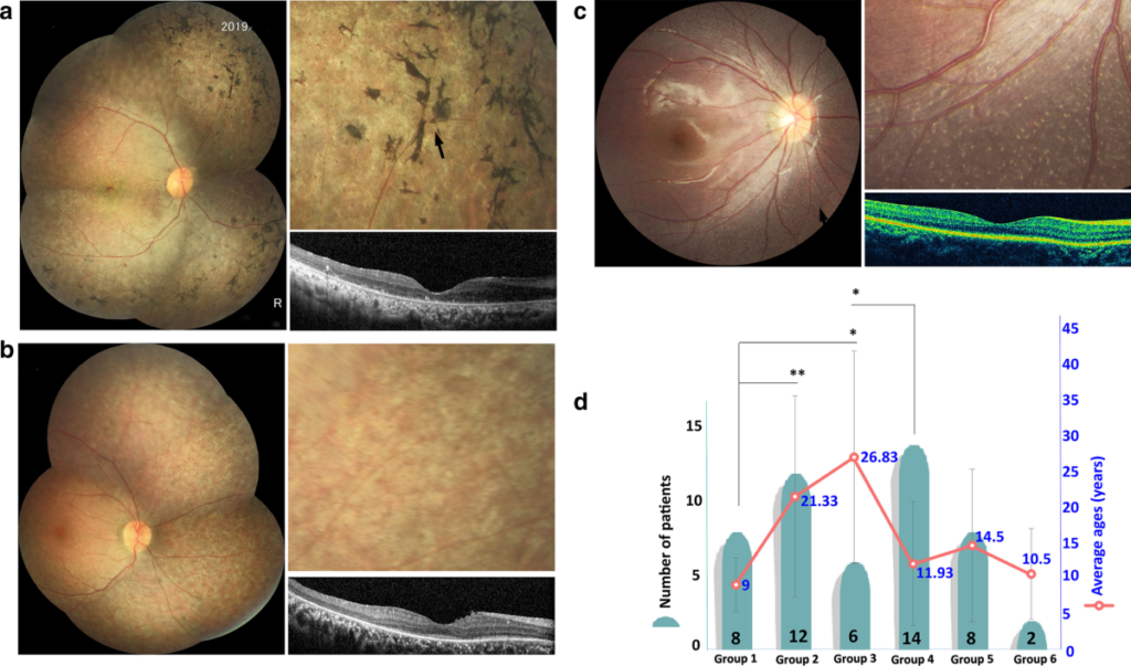
A thorough ophthalmic examination often reveals characteristic signs of retinal dystrophy, such as retinal pigment deposits, bone spicule pigmentation, and macular changes. Fundus photography and optical coherence tomography (OCT) can help document the extent of retinal damage.
3. Electroretinography (ERG)
Electroretinography (ERG) measures the electrical responses of the retina to light stimuli and is often used to assess the function of photoreceptor cells. In patients with biallelic RPE65 mutations, ERG typically shows reduced or absent responses, indicating impaired retinal function.
4. Visual Field Testing
Visual field tests help assess the extent of vision loss, particularly peripheral vision, which is often affected early in the disease.
Treatment Options
There is currently no cure for biallelic RPE65 mutation-associated retinal dystrophy, but significant progress has been made in developing gene therapies aimed at restoring normal RPE65 gene function. These therapies have shown promising results in clinical trials and offer hope for patients with this condition.
1. Gene Therapy: Luxturna (voretigene neparvovec)
Luxturna, a gene therapy developed by Spark Therapeutics, has been approved for the treatment of biallelic RPE65 mutations. This therapy involves the delivery of a functional copy of the RPE65 gene to the retina using an adeno-associated virus (AAV) vector. By restoring the missing or nonfunctional RPE65 enzyme, Luxturna aims to slow down or even halt retinal degeneration and improve vision.
In clinical trials, Luxturna has demonstrated improved visual acuity, enhanced light sensitivity, and expanded visual field in many patients with biallelic RPE65 mutations. However, it is most effective when administered early in the disease process, before significant retinal damage occurs.
2. Retinal Prosthetics
For advanced cases where gene therapy is not feasible, retinal prosthetics such as the Argus II Retinal Prosthesis System may offer some benefit. These devices help restore limited vision by using a camera to capture images and transmit them to the retina via a small implant.
3. Supportive Care and Low Vision Aids
Although there is no cure, supportive care such as the use of low vision aids, including magnifiers and specialized lighting, can help patients manage daily activities. Vision rehabilitation programs can also teach adaptive strategies to cope with vision loss.
4. Emerging Therapies
Ongoing research into gene editing techniques, such as CRISPR-Cas9, may offer future potential for more precise genetic modifications. Additionally, pharmacological approaches to support retinal health are also under investigation.
The Future of Biallelic RPE65 Mutation-Associated Retinal Dystrophy Treatment
As advancements in gene therapy, genetic research, and retinal implants continue, the outlook for patients with biallelic RPE65 mutation-associated retinal dystrophy continues to improve. Early diagnosis, coupled with cutting-edge therapies, offers the potential to preserve vision and enhance quality of life for affected individuals.

