Onychomycosis, medically referred to as tinea unguium, is a fungal infection of the nail unit caused predominantly by dermatophytes. These pathogenic fungi invade the nail plate, nail bed, and surrounding tissue, leading to discoloration, thickening, and eventual destruction of the nail. Onychomycosis due to dermatophyte is the most common cause of nail fungal disease globally, accounting for up to 90% of toenail infections.
The condition significantly affects quality of life, particularly among elderly, diabetics, and immunocompromised patients, and poses a cosmetic and functional concern. Prompt identification and appropriate antifungal treatment are critical to halting disease progression and preventing recurrence.
Etiology: Dermatophytes Causing Onychomycosis
The primary dermatophytes implicated in onychomycosis are:
- Trichophyton rubrum (most prevalent worldwide)
- Trichophyton interdigitale
- Epidermophyton floccosum
- Microsporum species (rarely)
These fungi exhibit a keratinophilic nature, allowing them to digest and inhabit the keratin-rich tissues of the nail unit. Dermatophyte nail infections are often chronic and progressive, with high recurrence rates if left inadequately treated.
Transmission and Risk Factors
Modes of Transmission
- Direct contact with infected individuals
- Contaminated surfaces (public showers, locker rooms, nail salons)
- Shared footwear or nail tools
Predisposing Risk Factors
- Age over 60
- Tinea pedis (athlete’s foot)
- Diabetes mellitus
- Peripheral vascular disease
- Immunosuppression (HIV, chemotherapy)
- Nail trauma
- Poor foot hygiene
- Occlusive footwear
Classification and Types of Dermatophyte Onychomycosis
There are five major clinical types based on the route and extent of fungal invasion:
1. Distal Lateral Subungual Onychomycosis (DLSO)
- Most common type
- Starts at the hyponychium or lateral nail fold
- Nail becomes discolored, thickened, and friable
2. White Superficial Onychomycosis (WSO)
- Fungal invasion confined to the dorsal nail plate
- Presents as chalky white spots on the surface
3. Proximal Subungual Onychomycosis (PSO)
- Rare, often associated with immunosuppression
- Fungus enters via the proximal nail fold
4. Endonyx Onychomycosis
- Fungus invades the nail plate without nail bed involvement
- Nail appears milky white but remains intact
5. Total Dystrophic Onychomycosis
- Final stage of any form
- Complete nail destruction, thickening, crumbling, and loss of nail
Pathogenesis of Dermatophyte Nail Infection
The infection process begins with fungal spores penetrating the nail bed or nail plate. Dermatophytes produce keratinases, enabling them to degrade keratin and colonize deeper layers of the nail.
Clinical Features and Symptoms
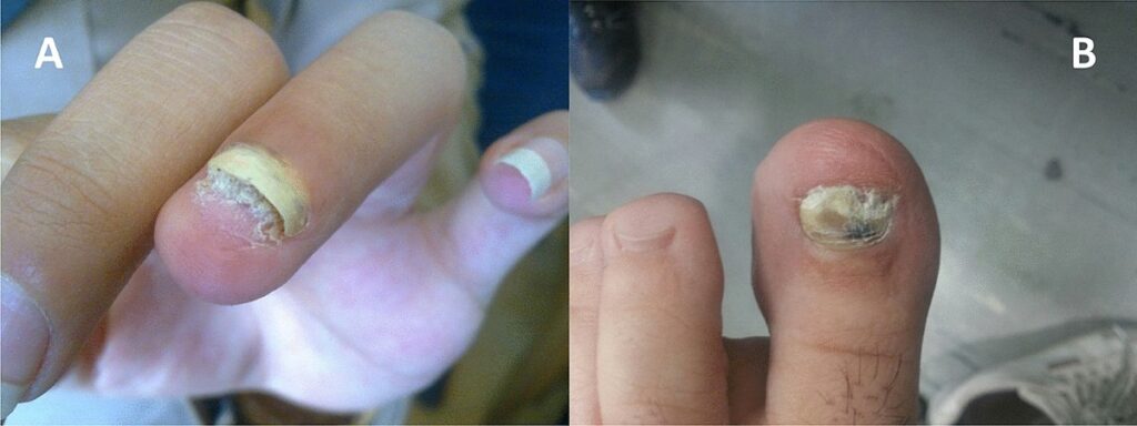
Visual Signs:
- Yellow, brown, or white nail discoloration
- Nail plate thickening
- Brittle, crumbly, or ragged nails
- Subungual debris
- Nail detachment (onycholysis)
Symptoms:
- Often painless
- Discomfort when walking (toenails)
- Social embarrassment due to unsightly nails
- May be secondary to tinea pedis or manuum
Diagnostic Methods for Dermatophyte Onychomycosis
Accurate diagnosis is essential to distinguish dermatophyte onychomycosis from other nail disorders such as psoriasis, lichen planus, or trauma.
Laboratory Tests:
1. Direct Microscopy (KOH preparation)
- Nail clippings examined after potassium hydroxide (KOH) treatment
- Detects fungal elements (hyphae)
2. Fungal Culture
- Confirms causative species
- Takes 2–3 weeks
- Essential for antifungal sensitivity testing
3. Periodic Acid-Schiff (PAS) Stain
- Performed on nail biopsy
- High sensitivity in detecting fungi
4. Polymerase Chain Reaction (PCR)
- Rapid, species-specific
- Detects fungal DNA in nail samples
Treatment of Dermatophyte-Induced Onychomycosis
Systemic Antifungal Therapy
1. Terbinafine
- First-line treatment for dermatophyte infections
- Dosage: 250 mg/day for 6 weeks (fingernails), 12 weeks (toenails)
- Fungicidal against Trichophyton spp.
2. Itraconazole
- Broad-spectrum antifungal
- Continuous or pulse dosing:
- Continuous: 200 mg/day
- Pulse: 200 mg twice daily for 1 week/month
- Useful for mixed fungal infections
3. Fluconazole
- Alternative in patients intolerant to others
- Dosage: 150–300 mg once weekly for 6–12 months
Topical Antifungal Agents
- Efinaconazole 10% solution
- Tavaborole 5% solution
- Ciclopirox 8% lacquer
- Ideal for mild to moderate cases or in combination with oral therapy
Adjunctive Measures
- Mechanical or chemical debridement
- Laser therapy (limited efficacy evidence)
- Photodynamic therapy (experimental)
Prevention and Recurrence Control
Preventive Strategies:
- Keep feet clean and dry
- Avoid sharing nail clippers or footwear
- Use antifungal powders in shoes
- Change socks regularly
- Trim nails straight and keep them short
- Disinfect pedicure tools
Recurrence Management:
- Monitor high-risk patients regularly
- Continue antifungal prophylaxis in diabetic or immunocompromised individuals
- Treat concomitant tinea pedis to avoid reinfection
Complications of Untreated Dermatophyte Onychomycosis
- Permanent nail deformity
- Secondary bacterial infections
- Cellulitis in diabetics or elderly
- Pain and gait abnormalities
- Psychosocial impact
Frequently Asked Questions
Q1: What causes onychomycosis due to dermatophytes?
It is caused by dermatophyte fungi, primarily Trichophyton rubrum, which invade and degrade nail keratin.
Q2: Can onychomycosis spread to other people?
Yes, the infection is contagious and can spread through shared surfaces or tools.
Q3: How long does treatment take?
Oral antifungals typically require 6–12 weeks for fingernails and 12–24 weeks for toenails.
Q4: Is topical treatment alone effective?
Topical agents work best in early or mild cases and may require long-term application.
Q5: Can the infection recur?
Yes, recurrence is common without proper foot hygiene or if associated conditions like tinea pedis are untreated.
Onychomycosis due to dermatophytes is a widespread and often neglected condition that requires a systematic diagnostic and therapeutic approach. Prompt recognition, appropriate use of oral and topical antifungals, and rigorous hygiene practices are crucial for achieving complete resolution and minimizing recurrence. As global awareness increases, early intervention and public education remain the cornerstones of effective disease management.