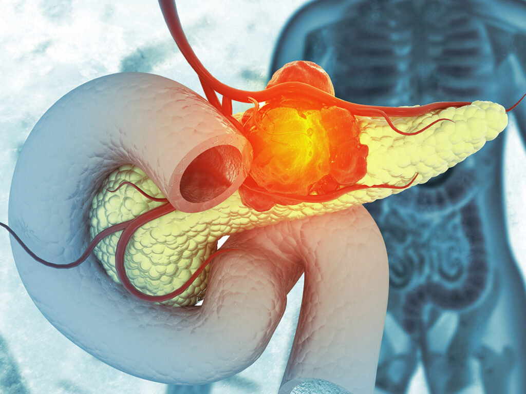
Nonfunctional neuroendocrine tumor: Nonfunctional neuroendocrine tumors (NETs) of gastrointestinal origin are a subset of rare neoplasms arising from neuroendocrine cells scattered throughout the gastrointestinal (GI) tract. Unlike their functional counterparts, these tumors do not secrete hormones in quantities sufficient to cause clinical symptoms of hormone excess. As a result, they are often diagnosed incidentally or during investigations for unrelated symptoms.
The most frequent sites of origin include the small intestine, rectum, stomach, and colon. Their incidence has been rising steadily, largely due to enhanced detection through endoscopy and imaging advancements.

Pathophysiology and Cellular Origin of GI Nonfunctional NETs
Nonfunctional NETs derive from enterochromaffin and enteroendocrine cells located within the mucosa and submucosa of the gastrointestinal lining. These tumors demonstrate neuroendocrine differentiation, evidenced by the expression of markers such as chromogranin A, synaptophysin, and CD56.
Tumor Development Mechanism
Mutations in tumor suppressor genes and dysregulation in cellular proliferation pathways lead to the development of well-differentiated or poorly differentiated NETs.
Common Sites of GI Nonfunctional NETs
| Location | Prevalence | Typical Characteristics |
|---|---|---|
| Small Intestine | High | Often multiple, submucosal nodules |
| Rectum | Moderate | Detected via endoscopy, often small |
| Stomach | Moderate | May arise from ECL cells |
| Colon | Less common | Often larger and more aggressive |
| Appendix | Rare | Usually found incidentally |
Clinical Presentation and Diagnostic Challenges
Symptoms of Nonfunctional NETs
Due to the lack of hormone-related symptoms, nonfunctional NETs are often clinically silent in early stages. When symptoms do occur, they are non-specific and depend on tumor size and location:
- Abdominal pain
- Change in bowel habits
- GI bleeding
- Bowel obstruction
- Palpable mass (advanced disease)
Diagnostic Approach
1. Imaging
- CT Scan / MRI: Assesses tumor size, invasion, and metastasis
- Somatostatin Receptor Imaging (Ga-68 DOTATATE PET/CT): Highly sensitive for NETs
2. Endoscopy and Biopsy
- Allows direct visualization and sampling
- Particularly effective for rectal and gastric NETs
3. Histopathology and Immunohistochemistry
- Expression of chromogranin A, synaptophysin, and Ki-67 index determines tumor grade
- Tumor grading per WHO classification:
| Grade | Mitotic Count | Ki-67 Index |
|---|---|---|
| G1 | <2 per 10 HPF | ≤2% |
| G2 | 2–20 per 10 HPF | 3–20% |
| G3 | >20 per 10 HPF | >20% |
Staging and Metastatic Spread
Staging follows the TNM classification (Tumor, Node, Metastasis), with common sites of metastasis including:
- Liver
- Regional lymph nodes
- Peritoneum
Advanced-stage NETs may present with multiple liver lesions or peritoneal carcinomatosis.
Management Strategies for GI Nonfunctional NETs
1. Surgical Resection
- Primary treatment for localized tumors
- May include segmental bowel resection, polypectomy, or radical surgery based on size and invasion
2. Medical Therapy
- Somatostatin analogs (e.g., octreotide, lanreotide) – used in advanced disease to stabilize progression
- Targeted therapies:
- Everolimus (mTOR inhibitor)
- Sunitinib (TKI for pancreatic NETs)
- Peptide Receptor Radionuclide Therapy (PRRT):
- Ga-68 DOTATATE PET-guided therapy using Lu-177-DOTATATE
3. Surveillance
- Low-grade tumors may be monitored with regular imaging and biomarker analysis
Prognosis and Survival Outcomes
Prognosis is highly dependent on:
- Tumor grade
- Stage at diagnosis
- Primary location
| Grade | 5-Year Survival Rate |
|---|---|
| G1 | >90% |
| G2 | 65–85% |
| G3 | <40% |
Early-stage, low-grade tumors have an excellent prognosis with curative surgical intervention.
Differentiating Functional vs. Nonfunctional NETs
| Characteristic | Functional NET | Nonfunctional NET |
|---|---|---|
| Hormone Production | Yes (serotonin, insulin, gastrin, etc.) | Minimal or none |
| Hormone-related Symptoms | Carcinoid syndrome, hypoglycemia, ulcers | Absent |
| Diagnosis | Symptom-based + labs | Often incidental or imaging-based |
| Management Complexity | Higher due to hormone control | Focused on tumor burden and metastasis |
Research Developments and Future Directions
- Biomarkers: Ongoing studies into circulating tumor DNA (ctDNA) and NETest for earlier detection
- Immunotherapy: Trials exploring PD-1/PD-L1 checkpoint inhibitors in high-grade neuroendocrine carcinomas
- Genetic profiling: Next-generation sequencing to personalize therapy and identify novel targets
- Artificial Intelligence: Enhancing endoscopic detection and histologic classification
Frequently Asked Questions:
What is a nonfunctional GI neuroendocrine tumor?
A nonfunctional GI neuroendocrine tumor is a neoplasm of the digestive tract that arises from neuroendocrine cells but does not produce hormones in clinically significant amounts.
How are nonfunctional NETs usually discovered?
They are often found incidentally during imaging, colonoscopy, or surgery for unrelated conditions due to their asymptomatic nature.
Are nonfunctional NETs curable?
Yes, if diagnosed early and confined locally, these tumors can often be cured surgically.
What is the role of Ki-67 in NETs?
Ki-67 is a proliferation marker used to grade NETs and predict their biological behavior and aggressiveness.
Can nonfunctional NETs become functional?
Transformation into a hormone-secreting tumor is extremely rare and not typically observed in clinical practice.
Nonfunctional neuroendocrine tumors of gastrointestinal origin represent a clinically silent but biologically diverse group of neoplasms. With advances in imaging, histologic grading, and molecular diagnostics, early detection and personalized management are becoming increasingly feasible. Continued research promises improved outcomes through targeted therapies and innovative diagnostic modalities.

