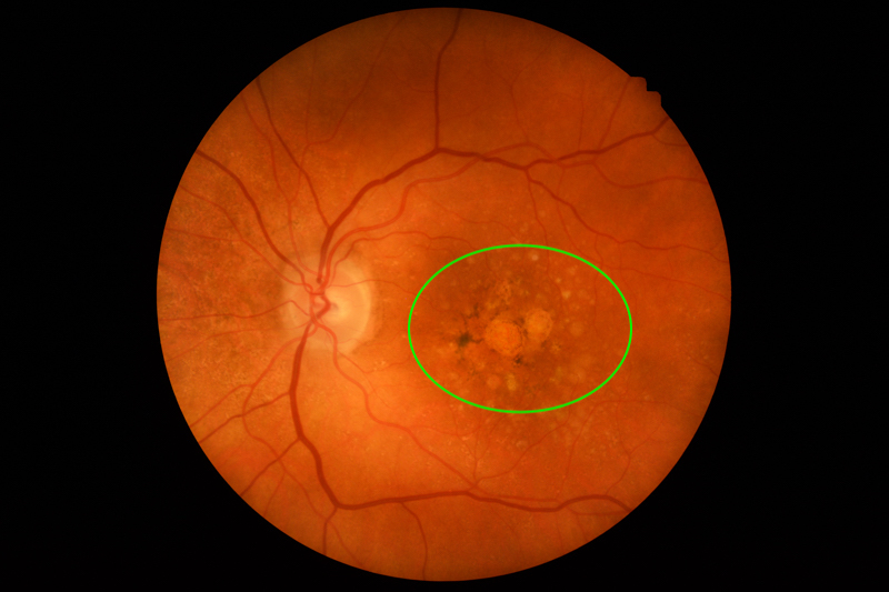
Neovascular age-related macular degeneration (nAMD), also known as wet AMD, is a progressive eye disease that primarily affects the macula, the central part of the retina responsible for sharp vision. It is characterized by abnormal blood vessel growth (choroidal neovascularization) beneath the retina, leading to fluid leakage, hemorrhage, and eventual vision loss. Unlike dry AMD, which progresses slowly, nAMD can cause rapid and severe central vision deterioration if left untreated.

Pathophysiology of nAMD
The development of neovascular AMD is driven by vascular endothelial growth factor (VEGF), which stimulates the formation of fragile and leaky blood vessels under the retina. These vessels disrupt normal retinal function, leading to:
- Macular edema (fluid accumulation)
- Hemorrhages within or under the retina
- Fibrotic scarring, which causes permanent vision impairment
Risk Factors for Neovascular AMD
Several factors contribute to the risk of developing nAMD, including:
Non-Modifiable Risk Factors
- Age: Predominantly affects individuals over 50
- Genetics: Variants in the CFH and ARMS2 genes increase susceptibility
- Family history of AMD
- Ethnicity: More common in Caucasians
Modifiable Risk Factors
- Smoking: Doubles the risk of AMD progression
- Hypertension: Contributes to vascular dysfunction
- Obesity and poor diet: Deficiencies in zinc, lutein, and omega-3 fatty acids elevate risk
- Excessive sunlight exposure: UV radiation may accelerate retinal damage
Symptoms of Neovascular AMD
Neovascular AMD presents sudden and noticeable changes in central vision, including:
- Blurred or distorted vision (metamorphopsia)
- Dark or empty spots (scotomas) in the central visual field
- Straight lines appearing wavy (Amsler grid distortion)
- Difficulty reading or recognizing faces
- Reduced contrast sensitivity and color perception
If untreated, vision loss can become irreversible, leading to severe impairment in daily activities.
Diagnosis of Neovascular AMD
1. Ophthalmic Examination
- Visual acuity test using the Snellen chart
- Amsler grid test to assess metamorphopsia
2. Advanced Imaging Techniques
- Optical Coherence Tomography (OCT): High-resolution imaging to detect fluid accumulation in the macula
- Fluorescein Angiography (FA): Identifies abnormal blood vessel growth and leakage
- Indocyanine Green Angiography (ICGA): Highlights deeper choroidal neovascularization
Regular screenings are crucial for early detection, especially in high-risk individuals.
Treatment Options for Neovascular AMD
1. Anti-VEGF Therapy (First-Line Treatment)
Anti-vascular endothelial growth factor (VEGF) agents block abnormal blood vessel formation and reduce leakage. These intravitreal injections have revolutionized nAMD treatment.
Common Anti-VEGF Agents:
| Drug Name | Mechanism | Injection Frequency |
|---|---|---|
| Ranibizumab (Lucentis) | VEGF-A inhibitor | Monthly or PRN |
| Aflibercept (Eylea) | VEGF-A and PlGF inhibitor | Every 8 weeks |
| Bevacizumab (Avastin) | Off-label VEGF inhibitor | Monthly |
| Faricimab (Vabysmo) | Dual VEGF and Ang-2 inhibitor | Extended intervals |
Regular injections are necessary to prevent disease progression and preserve vision.
2. Photodynamic Therapy (PDT)
- Uses verteporfin, a light-sensitive drug activated by laser, to target abnormal vessels.
- Typically used in persistent or refractory cases.
3. Laser Photocoagulation
- High-energy laser seals leaking vessels.
- Limited to extrafoveal lesions (away from the central macula) due to risk of scarring.
4. Emerging Therapies
- Gene therapy: Investigating RGX-314 and ADVM-022 for sustained VEGF suppression.
- Long-acting drug implants (Port Delivery System with ranibizumab).
- Stem cell therapy: Aims to regenerate damaged retinal cells.
Prognosis and Disease Management
Early intervention significantly improves visual outcomes. Patients receiving anti-VEGF therapy experience stabilization or improvement in vision in over 90% of cases.
Strategies for Long-Term Management
- Regular follow-ups with an ophthalmologist.
- Lifestyle modifications (smoking cessation, healthy diet).
- Nutritional supplements (AREDS2 formula) to support retinal health.
Visual Aids for AMD Patients
- Magnifying glasses and electronic reading devices
- High-contrast and large-print materials
- Adaptive lighting and color-enhancing lenses
Preventive Measures
While neovascular AMD cannot always be prevented, proactive steps can reduce risk and slow progression:
- Quit smoking – The most effective modifiable risk factor.
- Eat a macula-friendly diet – Rich in leafy greens, fish, and antioxidants.
- Manage cardiovascular risk factors – Control blood pressure and cholesterol.
- Wear UV-blocking sunglasses to protect against light-induced retinal damage.
- Regular eye exams – Essential for high-risk individuals over 50.
Neovascular age-related macular degeneration is a leading cause of severe vision impairment among older adults. Timely diagnosis and intervention with anti-VEGF therapy have transformed the prognosis, enabling many patients to maintain functional vision. Ongoing research into gene therapy, sustained drug delivery, and regenerative medicine offers hope for even more effective treatments in the future.
By understanding risk factors, symptoms, and treatment options, patients and caregivers can take proactive steps to manage this condition and preserve visual function.

