Kaposi’s sarcoma (KS) is a multifocal angioproliferative malignancy originating from lymphatic endothelial cells, characterized by the formation of vascular tumors primarily affecting the skin, mucous membranes, and visceral organs. First described by Moritz Kaposi in 1872, this neoplasm has been categorized into four distinct clinical variants: classic, endemic, epidemic (AIDS-associated), and iatrogenic (transplant-related).
Etiology and Pathogenesis
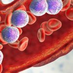
The etiological agent implicated in all forms of KS is human herpesvirus 8 (HHV-8), also known as Kaposi’s sarcoma-associated herpesvirus (KSHV). Transmission of HHV-8 occurs through various routes, including sexual contact, saliva, blood transfusions, and organ transplantation. While HHV-8 infection is necessary for the development of KS, it is not solely sufficient; the disease manifests predominantly in individuals with compromised immune systems, highlighting the role of immunosuppression in KS pathogenesis.
Clinical Variants
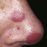
- Classic KS: Typically affects elderly men of Mediterranean or Eastern European descent. Lesions are usually confined to the lower extremities and progress slowly over years.
- Endemic KS: Occurs in sub-Saharan Africa, affecting both children and adults. It can present more aggressively, with lymph node involvement and visceral disease.
- Epidemic (AIDS-associated) KS: Associated with HIV infection, this variant is more aggressive and widespread, often involving the skin, oral mucosa, lymph nodes, and visceral organs.
- Iatrogenic (transplant-related) KS: Develops in organ transplant recipients undergoing immunosuppressive therapy. Lesions may regress upon modification or reduction of immunosuppressive treatment.
Clinical Presentation
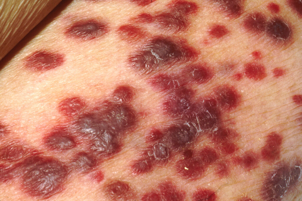
KS lesions typically present as painless, pigmented macules, papules, or nodules that may coalesce into plaques. These lesions are often red, purple, or brown due to their vascular nature. While cutaneous involvement is most common, KS can affect mucosal sites, lymph nodes, and visceral organs such as the gastrointestinal tract and lungs. Visceral involvement may lead to symptoms like gastrointestinal bleeding or respiratory distress.
Diagnosis
Definitive diagnosis of KS requires histopathological examination of biopsy specimens, revealing characteristic features such as spindle cells, neovascularization, and extravasated red blood cells. Immunohistochemical staining for HHV-8 latent nuclear antigen-1 (LNA-1) aids in confirming the diagnosis. Staging evaluations may include imaging studies and endoscopic procedures to assess the extent of visceral involvement.
Management Strategies
Treatment approaches for KS are tailored based on the variant, extent of disease, immune status, and overall health of the patient. Options include:
- Antiretroviral Therapy (ART): For epidemic KS, initiating or optimizing ART is crucial, as immune restoration can lead to lesion regression.
- Local Therapies: These include surgical excision, cryotherapy, and radiation therapy, suitable for limited cutaneous disease.
- Systemic Chemotherapy: Agents such as liposomal doxorubicin or paclitaxel are employed in cases with extensive cutaneous or visceral involvement.
- Immunomodulatory Agents: Interferon-alpha has shown efficacy in select cases, particularly in classic KS.
- Modification of Immunosuppression: In iatrogenic KS, reducing or altering immunosuppressive therapy can lead to lesion regression.
Prognosis
The prognosis of KS varies widely depending on the variant and extent of disease. Classic KS often follows an indolent course, while epidemic KS can be aggressive without effective ART. Early detection and appropriate management are essential to improve outcomes.
Kaposi’s sarcoma represents a spectrum of vascular neoplasms linked by HHV-8 infection and modulated by immune status. Advancements in understanding its pathogenesis and the development of targeted therapies have significantly improved patient outcomes. Ongoing research is essential to further elucidate the mechanisms underlying KS and to optimize therapeutic strategies.