Acute angle-closure glaucoma is a medical emergency that can lead to rapid vision loss if untreated. It occurs when the drainage angle in the eye becomes blocked, causing a sudden increase in intraocular pressure (IOP). This guide provides a detailed overview of acute angle-closure glaucoma, including symptoms, causes, diagnostic methods, and treatment options.

What is Acute Angle-Closure Glaucoma?
Acute angle-closure glaucoma (AACG) is a severe type of glaucoma characterized by a sudden rise in intraocular pressure. Unlike open-angle glaucoma, which develops slowly, AACG requires immediate medical intervention to prevent irreversible damage to the optic nerve and permanent vision loss.
How Does It Develop?
The condition occurs when the aqueous humor—a fluid inside the eye—fails to drain through the trabecular meshwork due to a blockage in the drainage angle. This blockage causes fluid buildup, leading to increased pressure within the eye.
Symptoms
Recognizing the symptoms of AACG is vital for early diagnosis and treatment. Common symptoms include:
- Severe Eye Pain: Intense pain in one or both eyes.
- Blurred Vision: Sudden loss of clarity or halos around lights.
- Headaches: Often localized around the affected eye.
- Nausea and Vomiting: Accompanying the eye pain and pressure.
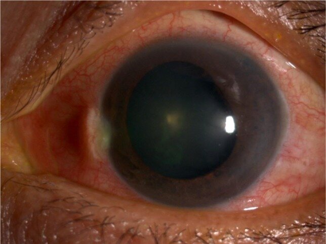
- Redness of the Eye: Often mistaken for conjunctivitis.
- Dilated Pupils: Pupils that react poorly to light.
These symptoms can develop rapidly, and prompt medical attention is essential.
Causes and Risk Factors
Understanding the causes and risk factors of acute angle-closure glaucoma helps identify individuals at higher risk.
Causes
- Blocked Drainage Angle: The primary cause of AACG.
- Iris Dysfunction: Forward displacement of the iris can obstruct the trabecular meshwork.
- Lens Thickening: Age-related changes in the lens can contribute to angle closure.
Risk Factors
- Age: More common in individuals over 50.
- Gender: Women are at higher risk than men.
- Ethnicity: Prevalent in East Asian and Inuit populations.
- Family History: Genetic predisposition increases the likelihood.
- Hyperopia: People with farsightedness often have narrower angles.
Diagnosis
Diagnosing AACG involves a thorough eye examination. Key diagnostic techniques include:
1. Tonometry
This test measures intraocular pressure. Elevated IOP is a hallmark of AACG.
2. Gonioscopy
A specialized lens is used to examine the drainage angle and confirm the blockage.
3. Ophthalmoscopy
The optic nerve is inspected for signs of damage caused by increased pressure.
4. Pachymetry
Measures corneal thickness, which can influence the diagnosis and management of glaucoma.
5. Visual Field Testing
Assesses peripheral vision loss, often affected in glaucoma.
Treatment Options
Immediate treatment is crucial to relieve intraocular pressure and prevent optic nerve damage. Treatment options include:
1. Medications
- Topical Beta-Blockers: Reduce aqueous humor production.
- Alpha Agonists: Lower IOP by reducing fluid production and increasing drainage.
- Oral Carbonic Anhydrase Inhibitors: Decrease intraocular pressure effectively.
- Hyperosmotic Agents: Used in emergencies to rapidly lower IOP.
2. Laser Therapy
- Laser Peripheral Iridotomy: A small hole is created in the iris to restore fluid flow.
- Laser Iridoplasty: Shrinks the peripheral iris to open the drainage angle.
3. Surgical Procedures
- Trabeculectomy: Removes a portion of the trabecular meshwork to improve drainage.
- Lens Extraction: For patients with lens-induced angle closure, cataract surgery may resolve the issue.
Long-Term Management and Prevention
Preventing acute angle-closure glaucoma involves regular eye check-ups, especially for individuals with risk factors. Key strategies include:
- Routine Eye Exams: Early detection of narrow angles or elevated IOP.
- Prophylactic Laser Iridotomy: Recommended for patients with anatomically narrow angles.
- Lifestyle Adjustments: Maintaining healthy habits to prevent general eye strain and conditions that may exacerbate angle closure.
Acute angle-closure glaucoma is a vision-threatening condition that demands prompt diagnosis and treatment. Awareness of its symptoms, risk factors, and prevention strategies can help individuals take timely action. Regular eye examinations and advanced treatments, including laser and surgical options, can mitigate risks and preserve vision.

