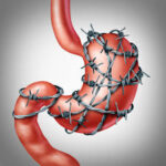
Gastroscopy, or upper gastrointestinal (GI) endoscopy, is a procedure used to visualize the esophagus, stomach, and duodenum. Adjunct techniques enhance diagnostic accuracy, improve procedural efficiency, and enable therapeutic interventions.
Types of Gastroscopy Adjunct Techniques
1. Endoscopic Ultrasound (EUS)
Endoscopic ultrasound combines high-frequency sound waves with endoscopy to produce detailed images of the GI tract and nearby structures. EUS is valuable for assessing submucosal lesions, staging malignancies, and guiding fine-needle aspiration (FNA).
2. Narrow-Band Imaging (NBI)
NBI enhances mucosal visualization by using specific light wavelengths to highlight vascular and tissue patterns. This technique improves the detection of early neoplastic changes in the esophagus, stomach, and duodenum.
3. Chromoendoscopy
Chromoendoscopy involves applying dyes such as methylene blue, indigo carmine, or Lugol’s iodine to better delineate mucosal abnormalities. It is particularly useful for identifying dysplastic or precancerous lesions.
4. Confocal Laser Endomicroscopy (CLE)
CLE allows real-time microscopic evaluation of the GI mucosa, reducing the need for biopsies. This technique is beneficial in diagnosing Barrett’s esophagus, gastric atrophy, and inflammatory bowel diseases.
5. Magnification Endoscopy
Magnification endoscopy provides high-resolution imaging of the GI mucosa, aiding in the identification of precancerous and cancerous lesions. It is often combined with NBI or chromoendoscopy.
6. Endoscopic Mucosal Resection (EMR) and Endoscopic Submucosal Dissection (ESD)
These advanced therapeutic techniques allow for the removal of precancerous and early-stage cancerous lesions, reducing the need for invasive surgery.
7. Capsule Endoscopy as an Adjunct to Gastroscopy
Capsule endoscopy, while primarily used for small bowel evaluation, can complement gastroscopy in detecting upper GI lesions in cases where conventional endoscopy is incomplete or inconclusive.
Importance of Gastroscopy Adjunct Techniques in Clinical Practice
Adjunct techniques enhance the sensitivity and specificity of gastroscopy, leading to improved early detection and better patient outcomes. These advancements play a crucial role in diagnosing and managing conditions such as:
- Esophageal and gastric cancers
- Gastroesophageal reflux disease (GERD) complications
- Barrett’s esophagus
- Peptic ulcer disease
- GI bleeding
- Helicobacter pylori infection
Challenges and Future Directions
While adjunct technologies significantly improve gastroscopy, challenges remain, including cost, accessibility, and the need for specialized training. Future advancements may focus on artificial intelligence (AI)-assisted diagnosis, improved endoscopic imaging, and robotic-assisted interventions.
Gastroscopy adjunct techniques have revolutionized the diagnosis and treatment of gastrointestinal disorders. As technology advances, these tools will continue to enhance early detection, therapeutic precision, and patient outcomes, making endoscopy a more effective and minimally invasive procedure.