Erysipelothrix endocarditis is a rare but serious form of bacterial endocarditis caused by Erysipelothrix rhusiopathiae, a gram-positive, facultatively anaerobic rod-shaped bacterium. This zoonotic pathogen primarily affects individuals with occupational exposure to animals, particularly swine, fish, and poultry. Due to its unique pathogenic mechanisms and resistance to common antibiotics, early diagnosis and targeted treatment are crucial for favorable outcomes.

Etiology and Risk Factors
Erysipelothrix rhusiopathiae is commonly found in soil, water, and animal products. Human infections usually occur through direct skin abrasions or mucosal exposure. The risk factors for Erysipelothrix endocarditis include:
- Occupational exposure (butchers, veterinarians, fishermen, farmers)
- Immunosuppression (diabetes, malignancies, cirrhosis)
- Prosthetic heart valves or pre-existing valvular disease
- Intravenous drug use (though rare for this pathogen)
Pathogenesis and Clinical Presentation
Once E. rhusiopathiae enters the bloodstream, it can colonize heart valves, leading to an inflammatory cascade. The infection manifests in two forms:
- Acute Endocarditis: Rapid onset with high fever, chills, and systemic toxicity.
- Subacute Endocarditis: Gradual progression with fatigue, weight loss, and low-grade fever.
Common symptoms include:
- Fever and chills
- Night sweats
- Fatigue and malaise
- Arthralgia and myalgia
- Heart murmur (new or changing)
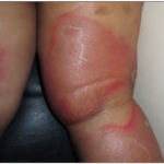
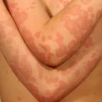
- Splinter hemorrhages, Osler nodes, and Janeway lesions (in some cases)
Severe complications can arise, such as embolic events (stroke, renal infarction), heart failure, and abscess formation.
Diagnosis
Due to its rarity, Erysipelothrix endocarditis is often misdiagnosed. A comprehensive diagnostic approach includes:
1. Blood Cultures
- E. rhusiopathiae grows in blood cultures but may require prolonged incubation.
- Gram stain: thin, pleomorphic gram-positive rods
2. Echocardiography
- Transthoracic echocardiography (TTE): May detect large vegetations
- Transesophageal echocardiography (TEE): More sensitive for prosthetic valve involvement
3. Histopathology and PCR
- Vegetation biopsy (if surgery is performed) may confirm the diagnosis.
- PCR assays can help in culture-negative cases.
Treatment Strategies
Effective management involves prolonged antibiotic therapy and, in some cases, surgical intervention.
1. Antibiotic Therapy
E. rhusiopathiae is intrinsically resistant to vancomycin, making beta-lactams the preferred treatment:
- First-line: Penicillin G or ceftriaxone (4-6 weeks)
- Alternative: Ampicillin or imipenem
- Penicillin allergy: Doxycycline or fluoroquinolones (limited data on efficacy)
For severe cases, combination therapy with gentamicin during the first 2 weeks may enhance bacterial clearance.
2. Surgical Intervention
Surgery is indicated for:
- Large vegetations (>10 mm) with embolic risk
- Heart failure due to valvular dysfunction
- Persistent bacteremia despite antibiotics
- Prosthetic valve endocarditis
Prognosis and Prevention
With early and appropriate treatment, mortality rates range between 30-40%, but delayed diagnosis increases the risk of complications. Preventive measures include:
- Proper wound care after animal handling
- Use of protective gloves and equipment
- Early recognition and treatment of E. rhusiopathiae infections to prevent hematogenous spread