Dyschromia refers to any alteration in the color of the skin or nails, encompassing both hyperpigmentation (darkening) and hypopigmentation (lightening). This condition can manifest due to various factors, including genetic predispositions, environmental exposures, and underlying medical conditions. Understanding the nuances of dyschromia is essential for effective diagnosis and treatment.

Types of Dyschromia
- HyperpigmentationHyperpigmentation is characterized by an increase in melanin production, leading to darker skin patches. Common forms include:
- Melasma: Often appearing as brown or gray-brown patches on the face, melasma is frequently associated with hormonal changes and sun exposure.
- Post-Inflammatory Hyperpigmentation (PIH): Dark spots that develop following skin injuries or inflammations, such as acne, eczema, or psoriasis.
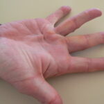
- HypopigmentationHypopigmentation involves a reduction in melanin, resulting in lighter skin areas. Examples include:
- Vitiligo: An autoimmune disorder where melanocytes are destroyed, leading to white patches on the skin.
- Post-Inflammatory Hypopigmentation: Lightened skin areas following skin trauma or inflammatory conditions.
Causes of Dyschromia
The development of dyschromia can be attributed to several factors:
- Genetic Factors: Certain pigmentary disorders, such as vitiligo, have a hereditary component.
- Sun Exposure: Ultraviolet (UV) radiation stimulates melanin production, potentially leading to conditions like melasma and sunspots.
- Hormonal Changes: Fluctuations in hormones, especially during pregnancy or with the use of oral contraceptives, can trigger melasma.
- Skin Injuries: Trauma to the skin, including cuts, burns, or inflammatory conditions, can result in post-inflammatory hyperpigmentation or hypopigmentation.
Diagnosis
Accurate diagnosis of dyschromia involves:
- Clinical Examination: A thorough skin assessment by a dermatologist to identify the pattern and extent of discoloration.
- Wood’s Lamp Examination: Utilizing ultraviolet light to detect changes in pigmentation not visible to the naked eye.
- Skin Biopsy: In uncertain cases, a small skin sample may be taken for histopathological analysis.
Treatment Options
Management of dyschromia depends on the underlying cause and may include:
- Topical Agents:
- Hydroquinone: A skin-lightening agent that inhibits melanin production, commonly used for hyperpigmentation.
- Retinoids: Vitamin A derivatives that promote cell turnover and can help in reducing pigmentation.
- Corticosteroids: Reduce inflammation and may be used in combination with other agents for certain types of dyschromia.
- Procedural Interventions:
- Chemical Peels: Application of chemical solutions to exfoliate the skin, promoting the growth of new, evenly pigmented skin.
- Laser Therapy: Utilizes specific wavelengths of light to target and break down pigmented cells.
- Microdermabrasion: A mechanical exfoliation technique that removes the outermost skin layer, aiding in the improvement of pigmentation irregularities.
- Sun Protection: Regular use of broad-spectrum sunscreens to prevent exacerbation of pigmentation disorders and protect against UV radiation.
Prevention Strategies
To minimize the risk of developing dyschromia:
- Sun Avoidance: Limit exposure to direct sunlight, especially during peak hours.
- Protective Clothing: Wear hats, long sleeves, and other protective garments when outdoors.
- Regular Skin Care: Maintain a consistent skincare routine using products suitable for your skin type