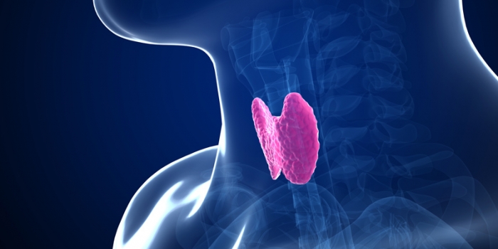
Differentiated thyroid carcinoma (DTC) is the most common type of thyroid malignancy, originating from follicular thyroid cells. It comprises two primary subtypes: papillary thyroid carcinoma (PTC) and follicular thyroid carcinoma (FTC). Both exhibit relatively slow growth and favorable prognoses when diagnosed early. This article provides an in-depth exploration of DTC, covering its classification, risk factors, diagnostic techniques, treatment modalities, and long-term management strategies.

Types of Differentiated Thyroid Carcinoma
Papillary Thyroid Carcinoma (PTC)
- Accounts for approximately 80% of all thyroid cancers.
- Often presents as a single or multifocal nodule in the thyroid.
- Frequently metastasizes to regional lymph nodes but retains an excellent prognosis.
- Commonly associated with BRAF and RET/PTC genetic mutations.
Follicular Thyroid Carcinoma (FTC)
- Represents 10–20% of thyroid cancers.
- More prevalent in iodine-deficient regions.
- More likely to spread hematogenously, particularly to the lungs and bones.
- Generally lacks lymph node involvement but has a slightly worse prognosis than PTC.
Risk Factors and Pathogenesis
Radiation Exposure
- Ionizing radiation exposure, especially during childhood, is a major risk factor.
- Radiation therapy for benign conditions (e.g., acne, enlarged tonsils) in the past has been linked to increased incidence.
Genetic Predisposition
- Familial clustering of DTC suggests a hereditary component.
- Genetic mutations such as RET, BRAF, and RAS are commonly implicated.
Iodine Intake
- Both iodine deficiency and excess play roles in thyroid carcinogenesis.
- FTC is more commonly associated with iodine-deficient diets.
Clinical Presentation
DTC often presents as a painless, palpable thyroid nodule. Additional symptoms may include:
- Hoarseness due to recurrent laryngeal nerve involvement.
- Dysphagia in cases of large tumors compressing the esophagus.
- Cervical lymphadenopathy in metastatic PTC.
While many cases are asymptomatic, incidental findings during imaging studies frequently lead to early detection.
Diagnostic Approach
Ultrasonography
- The first-line imaging modality for evaluating thyroid nodules.
- Identifies suspicious features such as microcalcifications, irregular margins, and increased vascularity.
Fine-Needle Aspiration Cytology (FNAC)
- Essential for distinguishing benign from malignant thyroid nodules.
- The Bethesda System categorizes cytological findings to guide clinical decisions.
Molecular Testing
- Detects genetic mutations (e.g., BRAF, RAS, RET/PTC) that assist in risk stratification and treatment planning.
Staging and Prognosis
DTC staging follows the TNM classification system, assessing:
- Tumor size (T)
- Lymph node involvement (N)
- Distant metastasis (M)
Prognostic Factors:
- Age: Patients <55 years generally have an excellent prognosis.
- Tumor size and extrathyroidal extension impact disease outcomes.
- The presence of distant metastases reduces survival rates but remains treatable.
Treatment Strategies
Surgical Management
- Total Thyroidectomy: Standard approach for most DTC cases to remove all thyroid tissue.
- Lobectomy: Considered in low-risk, small, unifocal tumors without metastases.
Radioactive Iodine (RAI) Therapy
- Administered postoperatively to ablate residual thyroid tissue.
- Particularly beneficial in cases with lymph node or distant metastases.
Thyroid-Stimulating Hormone (TSH) Suppression
- Levothyroxine therapy is prescribed to suppress TSH, reducing the stimulus for cancer cell growth.
Targeted Therapy
- In cases of RAI-refractory DTC, tyrosine kinase inhibitors (e.g., sorafenib, lenvatinib) are employed.
Long-Term Surveillance and Follow-Up
Thyroglobulin Monitoring
- Used as a tumor marker for disease recurrence.
- Rising levels post-treatment indicate potential residual or metastatic disease.
Neck Ultrasound
- Regular follow-up imaging to assess for recurrence or residual nodules.
Whole-Body Scan (WBS)
- Used selectively in patients receiving RAI therapy to detect distant metastases.

