Diagnostic test for pheochromocytoma is a rare neuroendocrine tumor arising from chromaffin cells of the adrenal medulla, causing excessive secretion of catecholamines such as epinephrine and norepinephrine. While hypertension is the most common clinical feature, severe insulin resistance is increasingly recognized as a significant metabolic complication in some patients. Elevated catecholamines interfere with insulin signaling, leading to impaired glucose metabolism, which can manifest as hyperglycemia or diabetes. Diagnosing pheochromocytoma in individuals with insulin resistance requires a structured approach incorporating biochemical, imaging, and genetic testing.

Biochemical Testing for Pheochromocytoma
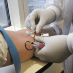
The first step in diagnosis involves biochemical confirmation of excessive catecholamine secretion. The preferred diagnostic tests include:
1. Plasma Free Metanephrines
- Most sensitive test for pheochromocytoma detection.
- Measures the breakdown products of catecholamines (metanephrine and normetanephrine).
- Requires fasting blood samples to minimize false positives.
2. 24-Hour Urinary Fractionated Metanephrines and Catecholamines
- Useful for confirming excess catecholamine secretion.
- Helps in cases where plasma metanephrines are inconclusive.
3. Clonidine Suppression Test
- Administered when plasma metanephrines yield borderline results.
- Clonidine, an α2-adrenergic agonist, suppresses catecholamine release in normal individuals but not in those with pheochromocytoma.
4. Glucose and Insulin Metabolism Markers
- Elevated fasting glucose and impaired glucose tolerance often accompany pheochromocytoma.
- Hyperinsulinemia with reduced insulin sensitivity suggests catecholamine-induced metabolic dysfunction.
Imaging Studies for Tumor Localization
Once biochemical tests confirm pheochromocytoma, imaging studies are required to determine tumor location and assess metastasis risk.
1. Computed Tomography (CT) Scan
- First-line imaging modality due to its high sensitivity.
- Pheochromocytomas appear as well-defined adrenal masses with variable enhancement.
2. Magnetic Resonance Imaging (MRI)
- Preferred in patients with contraindications to CT contrast agents.
- Tumors exhibit high signal intensity on T2-weighted sequences.
3. Functional Imaging for Metastatic or Extra-Adrenal Disease
- 123I-MIBG (Metaiodobenzylguanidine) Scintigraphy: Identifies functional adrenal and extra-adrenal pheochromocytomas.
- 18F-FDG PET/CT (Fluorodeoxyglucose PET scan): Used for aggressive or metastatic cases.
- 68Ga-DOTATATE PET/CT: Superior for detecting neuroendocrine tumors, particularly in SDHB-mutated cases.
Genetic Testing and Syndromic Associations
Up to 35% of pheochromocytomas have a hereditary basis. Genetic testing is crucial in patients with:
- Family history of pheochromocytoma or paraganglioma.
- Bilateral adrenal tumors.
- Extra-adrenal or metastatic disease.
Common Genetic Mutations
- RET (Multiple Endocrine Neoplasia Type 2 – MEN2)
- VHL (Von Hippel-Lindau Syndrome)
- NF1 (Neurofibromatosis Type 1)
- SDHx (Succinate Dehydrogenase Mutations – SDHB, SDHD, etc.)
Identifying a genetic mutation aids in risk assessment, surveillance planning, and familial screening.
Insulin Resistance and Metabolic Impact of Pheochromocytoma
Catecholamine overproduction disrupts glucose metabolism through multiple mechanisms:
- α2-Adrenergic stimulation inhibits insulin release from pancreatic β-cells, reducing glucose uptake.
- β3-Adrenergic activation increases lipolysis, leading to elevated free fatty acids, which impair insulin signaling.
- β2-Adrenergic activation in the liver enhances gluconeogenesis, further elevating blood glucose levels.
- Post-surgical risk of hypoglycemia exists due to sudden normalization of catecholamine levels, requiring careful glycemic management.