Severe insulin resistance (SIR) is a complex condition characterized by the body’s diminished response to insulin, leading to significant metabolic disturbances. Accurate assessment and management of SIR are crucial for preventing complications such as type 2 diabetes and cardiovascular diseases. Advanced diagnostic imaging techniques play a pivotal role in evaluating the extent of insulin resistance and associated metabolic abnormalities.

Advanced Imaging Modalities in Severe Insulin Resistance
In the evaluation of severe insulin resistance, several imaging modalities have proven effective in assessing adipose tissue distribution, organ-specific fat accumulation, and related metabolic complications.
1. Magnetic Resonance Imaging (MRI)
MRI is a non-invasive imaging technique that provides detailed images of soft tissues without ionizing radiation. In the context of SIR, MRI is utilized to assess:
- Visceral and Subcutaneous Adipose Tissue: MRI can differentiate between visceral adipose tissue (VAT) and subcutaneous adipose tissue (SAT), offering precise measurements of fat distribution.
- Hepatic Steatosis: MRI techniques, such as proton density fat fraction (PDFF), allow for the quantification of liver fat content, aiding in the diagnosis of non-alcoholic fatty liver disease (NAFLD), which is commonly associated with insulin resistance.
- Pancreatic Fat Content: Elevated fat deposition in the pancreas, detectable via MRI, has been linked to beta-cell dysfunction and insulin resistance.
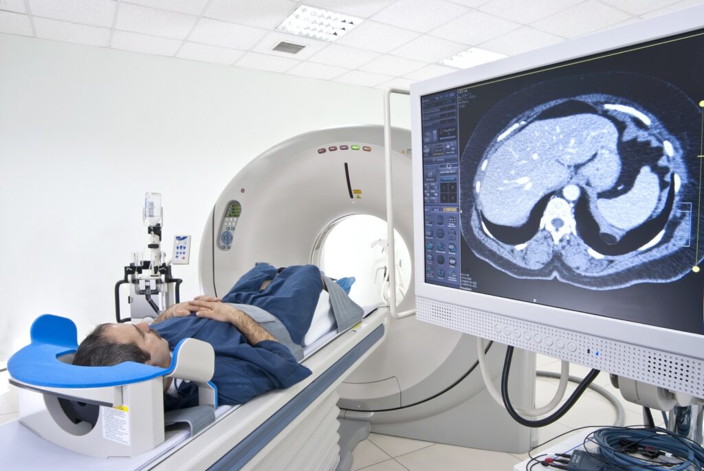
2. Computed Tomography (CT)
CT imaging is valuable in quantifying adipose tissue and assessing cardiovascular risk factors in SIR patients:
- Adipose Tissue Quantification: CT scans can accurately measure VAT and SAT areas, providing insights into fat distribution patterns.
- Coronary Artery Calcification: CT is instrumental in detecting calcified plaques in coronary arteries, serving as a marker for atherosclerosis, which is prevalent in individuals with insulin resistance.
3. Positron Emission Tomography (PET)
PET imaging, often combined with CT (PET/CT), offers metabolic insights:
- Glucose Metabolism Assessment: Using tracers like fluorodeoxyglucose (FDG), PET scans evaluate glucose uptake in tissues, highlighting areas of altered metabolism associated with insulin resistance.
4. Ultrasonography
While less detailed than MRI or CT, ultrasonography serves as a practical tool:
- Liver Assessment: Ultrasound can detect hepatic steatosis, providing a preliminary evaluation of liver fat content.
- Carotid Intima-Media Thickness (CIMT): Measurement of CIMT via ultrasound serves as a surrogate marker for atherosclerosis, offering insights into cardiovascular risk in SIR patients.
Integrating Imaging Findings into Clinical Practice
The integration of imaging findings with clinical and laboratory data enhances the comprehensive assessment of SIR:
- Risk Stratification: Imaging aids in identifying patients at higher risk for complications, facilitating targeted interventions.
- Monitoring Therapeutic Efficacy: Serial imaging studies can monitor changes in adipose tissue distribution and organ fat content, evaluating the effectiveness of therapeutic strategies.

