Deep vein thrombosis (DVT) and pulmonary embolism (PE) are critical components of venous thromboembolism (VTE), a condition with significant morbidity and mortality. DVT involves the formation of blood clots in deep veins, typically in the legs, while PE occurs when a portion of this clot dislodges and travels to the lungs, causing a potentially life-threatening blockage. Understanding the pathophysiology, risk factors, clinical manifestations, diagnostic approaches, and management strategies is essential for effective prevention and treatment.

Pathophysiology of Deep Vein Thrombosis and Pulmonary Embolism
The development of DVT is often explained by Virchow’s triad, which includes three primary factors:
- Venous Stasis: Reduced blood flow due to prolonged immobility or obstruction.
- Endothelial Injury: Damage to the blood vessel lining from trauma, surgery, or inflammation.
- Hypercoagulability: An increased tendency for blood to clot, which can be inherited or acquired.
When a thrombus forms in a deep vein, it can partially or completely obstruct blood flow. If a fragment of this clot breaks off, it can travel through the venous system to the pulmonary arteries, resulting in a PE. This embolism impedes blood flow in the lungs, leading to impaired gas exchange and increased strain on the right side of the heart.
Risk Factors
Several factors elevate the risk of developing DVT and PE:
- Prolonged Immobility: Extended periods of inactivity, such as long-haul flights or bed rest, can lead to venous stasis.
- Surgery and Trauma: Particularly orthopedic procedures, which may cause endothelial injury and immobilization.
- Malignancy: Certain cancers and their treatments can increase coagulability.
- Hormonal Factors: Use of oral contraceptives, hormone replacement therapy, and pregnancy can elevate risk.
- Genetic Predisposition: Inherited conditions like Factor V Leiden mutation enhance clotting propensity.
- Lifestyle Factors: Obesity, smoking, and dehydration contribute to risk.
Clinical Manifestations
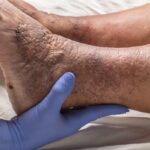
Deep Vein Thrombosis:
- Swelling: Typically unilateral leg swelling.
- Pain: Aching or cramping in the affected limb.
- Redness and Warmth: Localized erythema and increased temperature over the area.
Pulmonary Embolism:
- Dyspnea: Sudden onset of shortness of breath.
- Chest Pain: Sharp, pleuritic pain that may worsen with deep breaths.
- Hemoptysis: Coughing up blood.
- Tachycardia: Elevated heart rate.
- Syncope: Episodes of fainting or near-fainting.
It’s noteworthy that some individuals may remain asymptomatic or present with atypical symptoms, complicating diagnosis.
Diagnostic Approaches
Accurate diagnosis is crucial for effective management:
- D-Dimer Test: A blood test measuring fibrin degradation products; elevated levels suggest thrombosis but are not definitive.
- Compression Ultrasonography: Primary imaging modality for DVT detection, assessing vein compressibility.
- Computed Tomography Pulmonary Angiography (CTPA): Gold standard for PE diagnosis, providing detailed images of pulmonary vasculature.
- Ventilation-Perfusion (V/Q) Scan: Alternative imaging for patients contraindicated for CTPA.
Management Strategies
Treatment aims to prevent clot propagation, embolization, and recurrence:
- Anticoagulation Therapy: First-line treatment using agents such as low-molecular-weight heparin (LMWH), direct oral anticoagulants (DOACs), or warfarin.
- Thrombolytic Therapy: Considered in life-threatening PE or extensive DVT cases, involving clot dissolution with agents like tissue plasminogen activator (tPA).
- Inferior Vena Cava (IVC) Filters: Employed when anticoagulation is contraindicated, these devices prevent clots from reaching the lungs.
- Mechanical Thrombectomy: Physical removal of clots in select scenarios.
Duration of anticoagulation therapy is individualized, based on factors such as the presence of provoking factors, risk of recurrence, and bleeding risk.
Prevention
Preventive measures are vital, especially for high-risk individuals:
- Early Mobilization: Encouraging movement post-surgery or during long travel.
- Compression Stockings: Use of graduated compression stockings to enhance venous return.
- Pharmacologic Prophylaxis: Administration of anticoagulants in high-risk settings.
- Lifestyle Modifications: Maintaining a healthy weight, staying hydrated, and avoiding smoking.