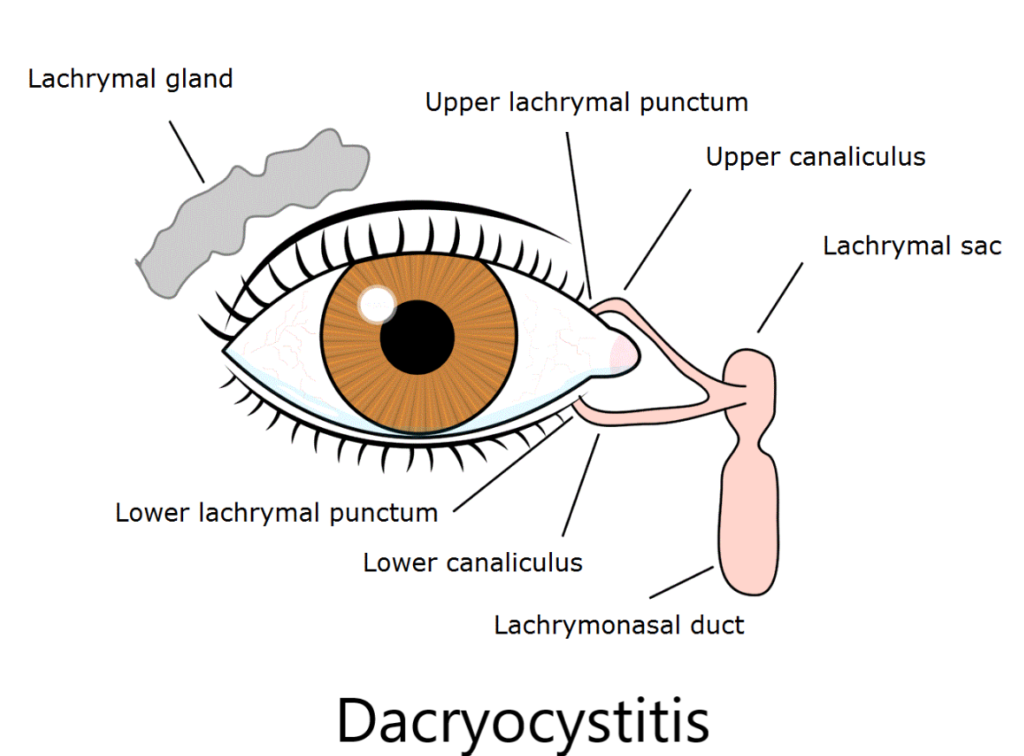
Dacryocystitis is an infection of the lacrimal sac caused by obstruction of the nasolacrimal duct. This blockage prevents normal tear drainage, leading to fluid accumulation, bacterial colonization, and inflammation. The condition can be acute or chronic, with varying severity and potential complications.

Causes and Risk Factors
Dacryocystitis primarily results from nasolacrimal duct obstruction (NLDO). Several factors contribute to this blockage:
- Congenital NLDO: Common in newborns due to incomplete duct development.
- Age-Related Changes: Narrowing of the duct with age increases susceptibility.
- Chronic Sinusitis or Nasal Conditions: Infections or structural abnormalities like nasal polyps can obstruct drainage.
- Trauma: Injury or previous surgeries can lead to scarring and obstruction.
- Tumors: Rarely, neoplasms can block the lacrimal system.
- Inflammatory Disorders: Conditions such as sarcoidosis or Wegener’s granulomatosis may contribute.
Symptoms and Clinical Presentation
Acute Dacryocystitis
- Severe pain and tenderness over the lacrimal sac region.
- Swelling and redness near the inner corner of the eye.
- Purulent discharge upon pressure application.
- Fever and malaise in severe cases.
Chronic Dacryocystitis
- Persistent tearing (epiphora).
- Mild swelling with occasional mucus discharge.
- Less pain and redness compared to acute cases.
Complications
If left untreated, dacryocystitis can lead to:
- Lacrimal abscess formation with risk of rupture.
- Preseptal or orbital cellulitis from infection spread.
- Fistula formation, creating an abnormal opening in the skin.
- Vision-threatening complications such as meningitis or cavernous sinus thrombosis.
Diagnosis
Dacryocystitis is diagnosed based on clinical examination and additional tests if needed:
- Physical Examination: Inspection and palpation of the lacrimal sac area.
- Fluorescein Dye Disappearance Test: Evaluates tear drainage efficiency.
- Microbiological Culture: Identifies the causative pathogen for targeted antibiotic therapy.
- Imaging Studies: Dacryocystography or CT scans are used in recurrent or complicated cases.
Treatment Approaches
Medical Management (Acute Dacryocystitis)
- Antibiotics: Broad-spectrum oral or IV antibiotics depending on severity.
- Warm Compresses: Alleviates pain and swelling.
- Pain Management: NSAIDs or analgesics as needed.
- Abscess Drainage: Incision and drainage if necessary.
Surgical Management (Chronic Dacryocystitis or Recurrent Cases)
- Dacryocystorhinostomy (DCR): The preferred surgical procedure to create a new drainage pathway for tears.
- Balloon Dacryoplasty: A minimally invasive technique using balloon dilation.
- Lacrimal Probing: Often used in congenital cases to open the duct.
Prevention and Prognosis
- Good eyelid hygiene and early management of eye infections.
- Timely intervention for nasolacrimal duct obstruction.
- Prognosis is generally excellent with appropriate treatment, though recurrence may require surgical intervention.

