Cutaneous Lupus Erythematosus (CLE) is an autoimmune skin disorder that presents with distinct dermatological symptoms. It can occur independently or in association with systemic lupus erythematosus (SLE). Understanding its types, symptoms, triggers, and treatment options is crucial for effective management.

Types of Cutaneous Lupus Erythematosus
CLE is classified into three major subtypes:
1. Chronic Cutaneous Lupus Erythematosus (CCLE)
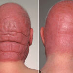
- Discoid Lupus Erythematosus (DLE): The most common form, characterized by scaly, disc-shaped lesions, often leading to scarring and pigment changes.
- Lupus Panniculitis: Involves deep dermal inflammation, resulting in firm nodules and tissue loss.
2. Subacute Cutaneous Lupus Erythematosus (SCLE)
- Non-scarring lesions that are photosensitive and often appear as annular or papulosquamous rashes.
- Frequently associated with anti-Ro/SSA antibodies and medication-induced lupus.
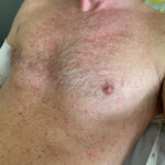
3. Acute Cutaneous Lupus Erythematosus (ACLE)
- Often manifests as the classic butterfly rash (malar rash) across the cheeks and nose.
- Strongly linked to systemic lupus erythematosus (SLE), commonly triggered by UV exposure.
Causes and Risk Factors
CLE arises due to immune system dysregulation, leading to an abnormal attack on skin cells. Key factors include:
- Genetics: HLA associations and familial predisposition.
- Environmental Triggers: UV radiation, infections, and exposure to certain medications (e.g., hydralazine, terbinafine, isoniazid).
- Hormonal Influence: Estrogen may contribute to higher prevalence in females.
- Immune Dysregulation: Autoantibody production (anti-Ro/SSA, anti-La/SSB, anti-dsDNA).
Symptoms and Clinical Presentation
- DLE: Scaly plaques, central scarring, hypopigmentation, or hyperpigmentation.
- SCLE: Red, ring-shaped or psoriasis-like lesions without scarring.
- ACLE: Malar rash, often exacerbated by sunlight exposure.
- Photosensitivity: Skin reactions triggered by UV exposure.
- Alopecia: Hair loss in affected areas, particularly in DLE.
Diagnosis of Cutaneous Lupus Erythematosus
Clinical Examination
A dermatologist evaluates lesion morphology, distribution, and associated systemic symptoms.
Laboratory Tests
- Autoantibody Screening: ANA, anti-Ro/SSA, anti-La/SSB, anti-dsDNA.
- Complement Levels: C3 and C4 depletion may indicate disease activity.
- Inflammatory Markers: ESR and CRP levels.
Skin Biopsy
- Histopathology: Interface dermatitis with lymphocytic infiltration.
- Direct Immunofluorescence: IgG and C3 deposition at the dermoepidermal junction.
Treatment and Management Strategies
1. Sun Protection and Lifestyle Adjustments
- Broad-spectrum sunscreen (SPF 50+)
- Protective clothing and UV avoidance.
- Smoking cessation (as smoking worsens CLE severity).
2. Topical and Systemic Medications
Topical Treatments
- Corticosteroids: Clobetasol, betamethasone (for active lesions).
- Calcineurin Inhibitors: Tacrolimus and pimecrolimus (for steroid-sparing effects).
Systemic Treatments
- Antimalarials: Hydroxychloroquine and chloroquine are first-line therapies.
- Immunosuppressants: Methotrexate, azathioprine, or mycophenolate mofetil for refractory cases.
- Biologics: Belimumab and rituximab (for severe, treatment-resistant cases).
3. Emerging Therapies
- JAK inhibitors and IFN-targeting agents are currently under investigation.
- mTOR inhibitors show promise in modulating immune response.
Prognosis and Long-Term Outlook
- DLE has a chronic course with potential for scarring.
- SCLE often resolves with treatment but may relapse.
- ACLE is typically associated with systemic lupus, requiring ongoing monitoring.
- Sun protection and adherence to treatment significantly improve outcomes.